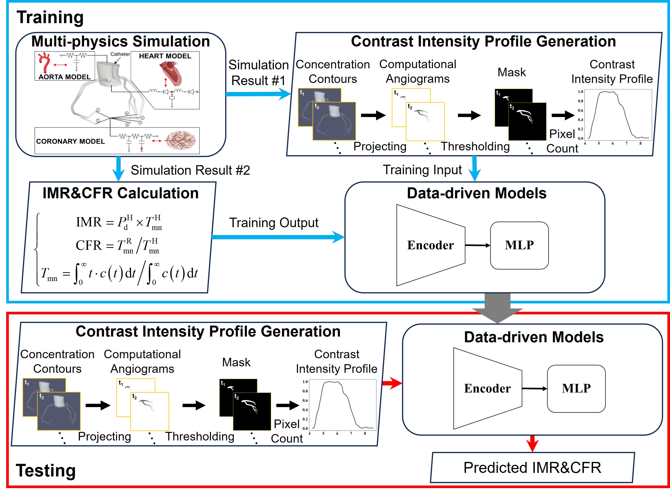
Coronary microvascular dysfunction (CMD), characterized by impaired regulation of blood flow in the coronary microcirculation, plays a key role in the pathogenesis of ischemic heart disease and is increasingly recognized as a contributor to adverse cardiovascular outcomes. Despite its clinical importance, CMD remains underdiagnosed due to the reliance on invasive procedures such as pressure wire-based measurements of the index of microcirculatory resistance (IMR) and coronary flow reserve (CFR), which are costly, time-consuming, and carry procedural risks. To date, no study has sought to quantify CMD indices using data-driven approaches while leveraging the rich information contained in coronary angiograms. To address these limitations, this study proposes a novel data-driven framework for inference of CMD indices based on coronary angiography. A physiologically validated multi-physics model was used to generate synthetic datasets for data-driven model training, consisting of CMD indices and computational angiograms with corresponding contrast intensity profiles (CIPs). Two neural network architectures were developed: a single-input-channel encoder-MLP model for IMR prediction and a dual-input-channel encoder-MLP model for CFR prediction, both incorporating epistemic uncertainty estimation to quantify prediction confidence. Results demonstrate that the data-driven models achieve high predictive accuracy when evaluated against physics-based synthetic datasets, and that the uncertainty estimates are positively correlated with prediction errors. Furthermore, the utility of CIPs as informative surrogates for coronary physiology is demonstrated, underscoring the potential of the proposed framework to enable accurate, real-time, image-based CMD assessment using routine angiography without the need for more invasive approaches.
More than 90% of the total flow resistance in the coronary circulation originates from pre-arterioles, arterioles, and capillaries within the microvasculature [1,2]. Consequently, microcirculation plays an important role in the regulation of coronary blood flow, ensuring a balance between oxygen and nutrient supply and metabolic demand. Coronary microvascular dysfunction (CMD), a significant concern in cardiology, is characterized by impaired blood flow and dysregulation within the microcirculation [2,3]. Structural abnormalities associated with CMD may arise from atherosclerosis, vascular remodeling, or fibrosis, whereas functional abnormalities can result from coronary vasospasm, endothelial cell and smooth muscle dysfunction, or metabolic derangements [4]. These abnormalities disrupt the delicate balance between myocardial perfusion and demand, leading to severe clinical outcomes, including heart failure, myocardial infarction, stroke, and increased mortality [5,6].
Diagnostic modalities to evaluate the functional state of CMD [7] include thrombolysis in myocardial infarction (TIMI) flow grade and TIMI frame count derived from coronary angiography, perfusion assessment using positron emission tomography (PET), and, less frequently, catheter-based techniques [8]. Coronary angiography, an X-ray-based imaging technique, is widely used in clinical practice, with millions of procedures performed annually worldwide. It provides high-resolution imaging of contrast agent dynamics within the coronary arteries, offering valuable insights into vascular morphology, motion, and deformation. TIMI flow grade and TIMI frame count are two angiography-based methods developed for CMD assessment, utilizing contrast washout patterns assessed visually by operators to evaluate microvascular perfusion and coronary flow dynamics [9]. Although these methods enable a rapid assessment of microvascular perfusion based on flow characteristics, they only provide qualitative and somewhat subjective and limited information, making them rarely used in practice. Myocardial positron emission tomography (PET) is an advanced imaging modality that provides high-resolution images and accurate measurements of myocardial blood flow at both rest and pharmacological stress, facilitating the detection of ischemia and microvascular dysfunction [10,11]. However, despite its diagnostic accuracy and prognostic value, the widespread adoption of PET remains limited due to high operational costs, the need for specialized equipment, and restricted availability to select medical centers.
Recently, invasive wire-based techniques, such as the index of microcirculatory resistance (IMR) and coronary flow reserve (CFR), have gained increasing attention as widely adopted invasive methods for assessing coronary microcirculatory function [12][13][14][15]. The assessment of these CMD indices involves the insertion of a coronary wire to simultaneously measure pressure and estimate flow. IMR is a quantitative measure of microvascular resistance, calculated using distal coronary pressure and hyperemic mean transit time to estimate the flow, providing an index of microvascular dysfunction independent of epicardial disease. CFR, on the other hand, represents the ratio of maximal hyperemic to resting coronary blood flow, reflecting both epicardial and microvascular contributions to coronary circulation. Notably, wire-based assessments have been shown to reliably predict myocardial viability following primary angioplasty for myocardial infarction and to determine the extent and severity of myocardial infarction in affected patients [16][17][18][19][20]. However, despite their clinical significance, these techniques remain underutilized due to their invasive nature, procedural complexity, and associated patient risks. Consequently, there is a pressing demand for a non-invasive computational approach to replicate these wire-based measurements.
Several studies have developed computational fluid dynamics (CFD)-based methods to compute the IMR [21][22][23] and CFR [24] using geometries reconstructed from angiographic data. However, these approaches face significant challenges related to the setup of boundary conditions in CFD models. In many cases, either invasive fractional flow reserve (FFR) is employed to provide pressure boundary conditions, or TIMI frame count is used to estimate flow boundary conditions. Invasive FFR, similar to invasive IMR or CFR assessment, is costly and poses additional risks to patients. On the other hand, flow estimation via TIMI frame count lacks accuracy. Moreover, the high computational cost associated with CFD simulations leads to prolonged processing times and increased demand for computational resources. Yong et al. introduced a novel method for IMR calculation in the presence of epicardial stenosis by establishing a linear regression model that relates coronary FFR to myocardial FFR [25]. However, this approach could be enhanced by employi
This content is AI-processed based on open access ArXiv data.