Restoring missing low scattering angle data in two-dimensional diffraction patterns of isolated molecules
Anisotropic two-dimensional diffraction signals contain more information than the conventional isotropic signals for both gas phase ultrafast electron and X-ray diffraction experiments and are common in typical time-resolved diffraction experiments d…
Authors: Yanwei Xiong, Martin Centurion
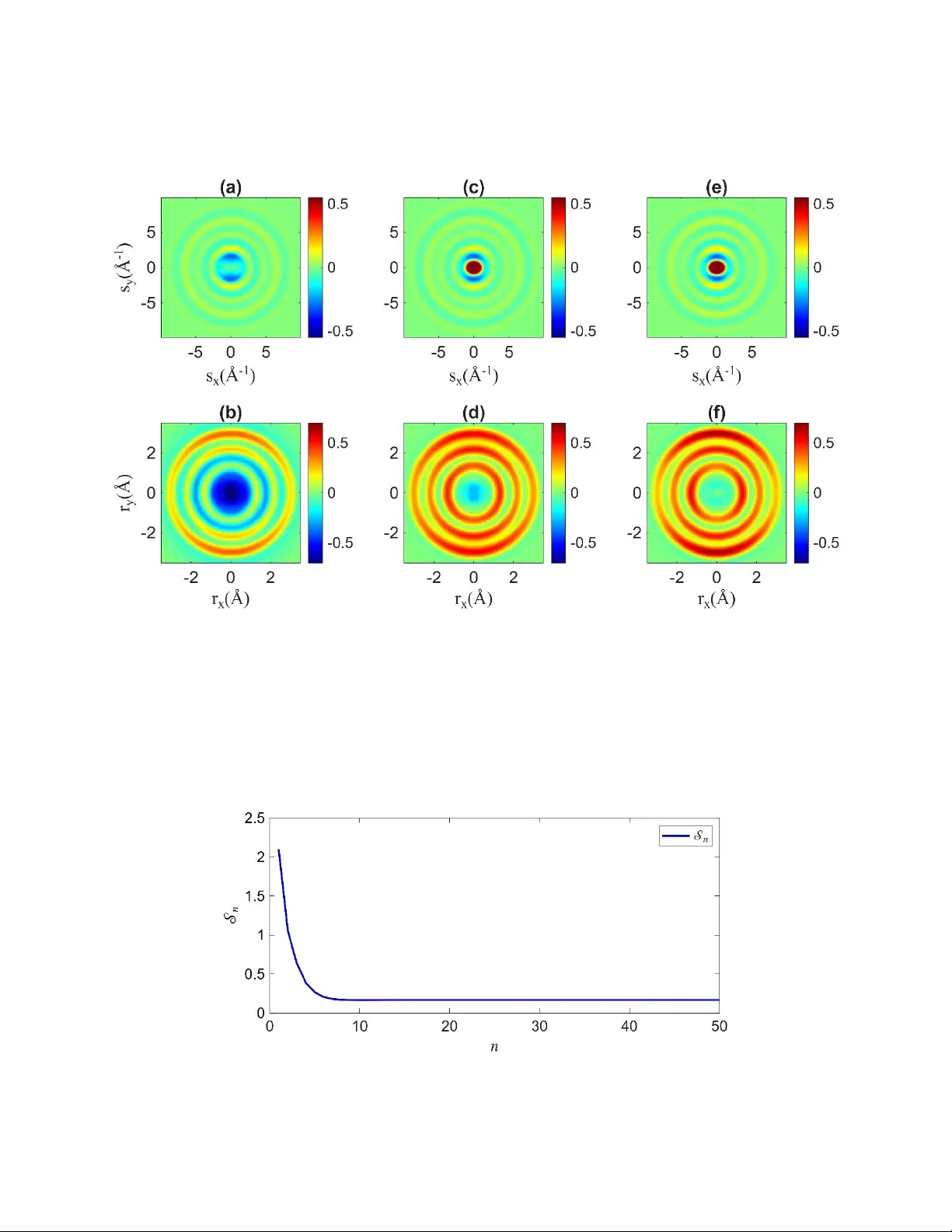
*Contact author: yxiong3@unl.edu † Contact author: martin.centurion@unl.edu 1 R e s t o r i n g m i s s i n g l o w s c a t t e r i n g a n g le d at a i n t w o - d i me n s i o n a l d i f f r a c t i o n pa t t e r n s o f i s o l a t e d m o l e c u l e s Yanwei Xiong * , Martin Ce nturion† Department of Phy sics and Astronomy, Univ ersity of Nebraska-Lincoln, Lincoln, N ebr aska, 68588, USA ABSTRA CT . Anisotropic tw o -dimensional diffraction signals contain more informatio n than the conventional isotropic signals for both gas phase ultrafast electron and X-ray diffraction experiments and are common in typical time-resolved diffraction experiments due to the use of linearly polarized lasers to excite the sample that imprint s sp atial anisotropy on the molecules. We report an i terative algorithm to restore the missing da t a at low scattering a ngles in a two-dimensional diffrac ti on signal, which is essent i al to obtain r eal-space representation . The iterative algorithm trans f orms two-dimen si onal s i gnals back and forth between the momen t um transfer doma i n and the real space doma i n through Fourier and A bel transforms and apply real space constraints to retrieve missing signal at low scattering angle s. The algorith m only requires an approximate a-priori knowledge of the shortest and longest internuclear distances i n the molecule. We demonstra ted successful retrieval of the missing signal in simulated patterns and in experimentally measured diffraction patterns from l aser-induced alignment of t r ifluoroiodo m ethane molecules. I. INTRODUCT IO N Gas-phase ultrafast electron diffraction (GUED) and ultrafast X -ray diffraction (UXRD) are potent techniques for ca pturing the structures and nuclear motions of isolated molecules during chemical reactions with exceptional femtosecond and sub -angstrom resolutions [1, 2]. In GUED and UXRD experiments, a pump laser excites the molecules, while a probe pulse, typically an electron pulse (tens of keV [3 -6] or MeV [7 -10] kinetic energy) or an X-ray pulse (around 10 keV energy) [11-14], interrogates the evolving molecular struc ture. The time delay betwe en the t wo pulses is adj usted to take a se ries of two-dimensiona l (2 -D) diffraction patterns, in which the info r mation of both the unexc it ed molecules and t he evolving structure after photoexcitation could be extracted t hrough real-space retrieval or comparison to theoretical predictions [15]. The small interaction cross-section and low density of molecules in a gas phase sample results in m ost incident electrons or photons passi ng s traight through the gas eous sample withou t i nteracting. A beam stop or ho l e is required to prevent t he direct transmitted b eam from striking the detector, which protects the detector from damage and i s crucial f or collecting signals of scattered electrons or photons. This leads to the l oss of diffraction signals at low scattering angles. I n addition, the maxi m um scattering angles available for t ypical diffraction experiments are li m ited by th e size of the d et ector or t he signal levels which decre as e rapidly with increasing scattering angle. Therefore, the accessible range of momentum transfer in typical diffraction experiments is limited by a minimum value and m aximum value . The value does not caus e issues in the r eal-space retrieval wh en treated appropriately, such as using a Gaussia n damping function. However, the si gnal at th e low scattering angles is es sential to o bt ain the real-spa ce pair distribution func tion (PDF). The analysis of GUED an d UXRD signals has been mostly r elegated to one-dimensional (1- D) in both momentum transfer domain and real space, which corresponds to the isotropic scattering signal. Multiple 2 methods have been developed to address t he issue of the missing isotropic signal at l ow scattering angles, such as filling the missing signal by smooth interpol at ion [16, 17], filling with the signal produced by simulations [3, 18-22], carrying out data analys i s in momentum tran sf er space [13, 23-27], r etrieval of structure with genetic algorithm [28-31], m odel-free inversion technique producing a super -resolved PDF from experiment al data [32], etc . Recently, we introduced an iterative algorithm in ref. [ 33] to restore the missing isotropic signal at small scattering angles, i .e. from 0 Å -1 t o , wit h the available experimental signal from to . The iterative algorithm is adapted from the ideas of existing phase retrieval algorithms [34-39], and iteratively transform s the data back and f orth between the m omentum t r ansfer and real spaces and applies a support constraint in real space, while allowing the missing signal from 0Å -1 to to be retrieved. Compared to the exist ing m ethods [3 , 13, 16-32], this m ethod has advant age s that it is simple to implemen t and requires only a minimal amount of a-priori knowledge of the molecule and can be applied to experi m ents with multiple reaction channels. In addition, this method could also be used to separate the inel astic scatte r ing from the elastic scattering in the GUED si gnals. While the analysis of diffraction signals has been mostly relegated to isotropic scattering signals, anisotropic 2-D diffraction patterns are very common in GUED and UXRD experiments since l inearly polarized laser pulses are commonly used to excite the molecules, which imprints a characteristic spatial anisotropy on the molecula r ensemble and results in an i sotropic patte rns [40]. The 2-D scatter i ng signal of a spatially anisotropic sample poss es ses m ore inf orm at ion t han the conventional isotropic signals. For example, 2-D GUED si gnals have been use d to retrieve additional i nformat i on about the dynamics of molecular reactions, i ncluding identifying bond distances and angles of molecules in the ground state [4, 41, 42], measurement of atom pair angular distributions [5, 43, 44], transient m olecula r structures [8, 23, 45] and dynamics [ 46] during la se r induced r eactions. The 1-D Fourier-sine transform is used to retrieve the real space representation of isotropic scattering signal, whereas the real space retrieval of anisotropic 2 -D diffraction patterns is more compl i cated and hinges on the 2- D Fourier and Ab el inversions. Therefore, t he it erative algorithm for i sotropic signal restoration cannot be used to retrieve the miss i ng da ta in a nisotropic 2-D diffraction pa tt erns. In this work, we develop the iterative algorithm to retrieve the missing signal at l ow scattering angles in 2-D diffraction signals by combining the idea of isot r opic signal restoration and the equa ti ons for real space retrieval from anisotropic 2-D diffraction patterns [5, 23, 42, 44]. We demonstrate the accuracy of the retrieval al gorithm with simulated and expe rimental electron scatte r ing signals of triflu or oiodome t hane (CF 3 I). II. THEORY In this section, we first review the elect r on scattering t heor y for gas phase molecules. We then describe the iterative algorithm that restores the inaccessib l e signal in electron scattering measur em ent with the available 2- D diffraction signa l . A. Electron scat t ering the ory The conventional theory of el ectron scattering from isolated molecules has been described in detail previously [ 19, 47-50] . The elastic scattering from a neutral m olecule c an be well approximated using the independent atom model, where the potential of ea ch atom that constituents molecule is assumed to be spherically sy mmetric and the bo nding effects bet ween a toms a re ignored [ 29, 47, 51 ]. I n electron scattering from isolated molecules, electron wave s s cattered fro m atoms w ithin the sa m e molecule interfere. The ela stic scattering i nt ensity of a si ngle molecu l e that consi sts of N atoms is given by 3 , (1) where is a vector pointing from the kth atom to the jth atom with length equal to the interatomic distance, and is the momentum tr ansfe r with magnitude , in which is the wavelength and i s the scattered angle o f electrons, and is the atomic scat tering amplitude of the jth a tom [52, 53]. The tot al e lastic diffrac t ion intensity from a gas phase samp le is an incoheren t sum of the scattering signals from al l the molecules i n the ens emble. Here we focus on 2-D di ffraction patterns produced by a sample o f molecules excited by a linearly polarized laser pul se . Th e tot al scattering intensity can be separated into atomic scattering intensity , which contains no information of the molecular structure, and the molecular scattering intensity , i n which molecular geometry information is enco ded [54, 55]. , (2) , (3) , (4) where are p ol ar and azimuthal angles that de scribe t he atom pair of a molecule in the lab fra m e, and is the angular distribution of atom pair . The general relation of the atom-pair angular d istribution and probability density of t he molec ul ar orientation, which is a func t ion of the Eu ler ang l es, is g iven by eqn (1) in [15, 44]. When the molecules are in a random spatial distribution, the atom-pair angular distributions are , and eqn (4) becomes Debye scattering equation, fo r mulated as , corresponding the isotropic scattering signal [54, 55]. In thi s case, the pattern consis ts of a series of concen tric rings that decrease rap i dly as s increases. How ever, i n general cases, the integra l in eqn (4 ) cannot be exp r essed as an analytical form like the Debye equation. The atomic scattering amplitudes decreases approximate l y as s -2 as s increases. W e t herefore define the resca led molecula r scattering intensity to remo ve the r apidly decreas i ng trend and highlight the oscillations at highe r mome nt um trans fers in the diffrac ti on pat tern, formulated as . The ato m - pair d i stance and angular distribution of all atom pairs in real space can be retrieved from the modified pair distribution function (MPDF), which i s produced b y the Fourier i nversion ( ), fol lowed by the Abe l inversion ( ), of , given by [5, 23, 44] , (5) where , si gnifies convolution, stands for correlation, is the ideal pair distribution function (PDF ) , and is the Fourier transform of the normalized atomic scattering amplitude . The MPDF can be understood as an angularly dependent pair distribution function. In time-resolved e xperiments, t he tim e -dependent signal can b e isolated using the diffrac tion-difference method, given by the difference of total scatte r ing: , (6) 4 where the variable denotes the t ime delay of electron pulse with respect t o the laser excitation, is the total scattering intensity at time ,which corresponds to a time before the arrival of the laser pulse, i.e. before the m olecules are excited [19, 42]. Corresponding ly, t he rescaled molecular scattering intensity can be defined as . The can be calculated by replacing in eqn (5) with . B. Retrieval method In this section, w e describe the algorithm t o restore the missing low- s signal in 2-D diffraction patterns . First, we establish t he transform pairs i n the signal and transform domains. Second, we mathematically model the typical 2- D experimental scattering pattern with a limited momentum transfer from to , and the artifact t hat appears in real space due to the missing signal. Third, we apply the constraint in real space that itera t ively reduc es t he ampli t ude of the art i fact. The rescaled molecular scattering intensity that is limited by the maximum momentum transfer can be defined as (7a) where . For the diffract ion -difference signal in t ime-resolved experiments, is given by (7b) where , are the newly pr oduce d interato mic d i stances and angu l ar distribut ions afte r lase r excitation, and , are t he interatomic distances and angular distributions before l aser excitation. The transform pai rs in signal and transform d omains are given by (8 a) (8b) Where is a damping function used to avoid artifacts due to the discont inuity of the signal at , and and ar e the forward Fourier and Abel transform. is the MPDF of rescaled scattering pattern with maxi m um momentum transfer , and can be written as as function o f polar coordinates in rea l space, where and . When the is given by eqn (7a), the is g i ven by , and is t he measured PDF, which is the convolution of and the Fourier inversion of the multiplication o f and the funct ion truncating the d iffraction signal due to the limited size of th e detector. Suppose the 2- D dif f raction signal from 0 Å -1 to is inaccessible and is filled with a first-guess function . The r escaled scattering intensity that corresponds t o the m easured diffraction signal can be defined as 5 (9) Similar to transform pairs in eqn (8a) and (8b), the transform pairs of the m easured signal with incorrect information in t he region can be defined as (10a) (10b) The can be written as , (1 1) where is the true signal that we are seeking, and is an artifact in r eal space due to the difference between and t he true signal in the low momentu m transfer from 0 Å -1 to . The can be expressed as . (1 2) Also, we have the following relation . (1 3) Our purpose of this work is to iteratively reduce t he amplitude of to retriev e using an a- priori k nowledge of the minimum an d maximum interatomic distances in the molecula r s t ructure. The conditions for successful retrieval are: (a) The momentum transfer of the available scattering pattern from to needs to be sufficiently large such that the approximate distribution of the atom-pair distances could be obtained. (b) The mome ntum transfer of the inaccessible signal 0 Å -1 to should be sufficiently small such that has a broad er distribution than that of . (c) An approximate a- priori knowledge o f t he minim um and maxim um interatomi c distance s of the mo lecule is known. Figure 1 shows a block diagram of the retrieval algorithm with iteration number denoted by n . The steps are as follows. (1) At the start, for n =1, we set . (2) A Fourier inversion ( ), followed by Abel inversion ( ), is applied to to generate . (3) The real-space support constraint f unction , based on a- prior knowledge of the molecule, is applied to truncate the artifact signal beyond t he minimum and maximum distances of the molecule to produce . The constraint function is a 2- D band-pas s filter that selects the sig nal within the range , given by , (14) where and , and is a positive integer. The sharpness of the filter is determined by . The is constrained within the constraint function , while extends beyond the co nstraint function. Therefo r e, the ampli t ude of the artifact will be red uced with each iteration. (4) The forward Abel transform ( ), fol l owed by the Fourier transform ( ), is applied to to generate . (5) We decompo se t he diffraction pattern into Legendre polynomials [40], formulated as , (1 5) 6 where is the azimuthal angle on the detector plane, and is the ith order Legendre polynomial . For molecules pumped by a linearly polarized lase r , only even orders are consi dered due to the cylindric al symmetry of the diffraction pattern. (6) The decomposed components of are . The and are stitched together to produce , in which t he signal from 0 t o is closer t o the true signal compared to the previous iteration. To avoid a discontinuity in , the is multiplied by a rescaling factor, given by / , where is a small value. (7) We genera te the new 2-D pattern using component s and eqn (15 ). ( 8) Repeat step (2) by replacing with to generate and , followed by steps (3)-(7 ). The retrieval er ror is defined as t he total sum of the square of the difference be tween and . . (1 6) According to eqn (13), approaches as approaches zero. The iteration is stopped when decreas es to a small nu mber and reaches a plateau. Figure 1. Block d iagram of the retrieval algorithm that restore the low - s signal in 2-D diffraction pattern. The iteration numb er is den ot ed by n . 7 III. TEST WITH A SIMU LATED PATTERN In this se ction, we test the retrieval algorithm using a calculated diffraction pattern of CF 3 I. The kinetic energy o f the e l ectrons used in the diffraction pattern ca lculation is 9 0 keV, and the scattering am plitude o f the atoms are tabulated in [56]. Since linearly polarized lasers are commonly used in GUED experiments, we consider the case of the probability density of the molecular orientation produced by a linearly polarized laser pulse, which imposes cylindrica l symmetry about the laser polariza ti on directi on. Here we use a simple probability density, given by , for the cal culation o f 2-D diffraction pattern. The other atom-pair angula r distributi ons can be obtained using t he method given in [1 5, 44]. The structure of a CF 3 I molecule [57] is shown in Figure 2(a), where the iodine at om is purple, the carbon is grey, and the fluorine atoms ar e green. The m inimum and m aximum i nteratomic distances of t he CF 3 I molecule are r CF = 1.33 Å and r FI =2.89 Å. We defined the 2-D constraint function with r 1 = 1.15 Å , r 2 = 3.20 Å and . The dampi ng constant is . We use eqns (4) and (7a) and the m ethod in [15] to calculate the and , and select the s -range 1.6 Å -1 10 Å -1 of the 2-D pattern as the available signal which matches the typ ical rang e in GUED measurement s [6, 9]. The pattern is decomposed into Legendre pol ynomials , where , and higher orders are ignored due to their negligible amplitudes (higher orders can be included in the Legendre projection if the amplitudes are not small). We use a line ar interpolat i on to fill the miss i ng region in from 0 Å -1 to 1.6 Å -1 , formulated a s . The and are shown as the solid black lines i n Figure 2 (a) and (b). The retrieved (see Figure 2 ) and 2-D di ffraction p atterns (see Figu r e 3) are obtained si m ultaneously with the iterative algorithm. The retri eved after 50 iterations are the solid blue lines, and t he true are dashed red lines, shown in Figure 2(a) -(b). The difference between and is significantly reduced, and the restored data from 0 to is i n good agreement with the true signal . Figure 2. The input and restored . (a) is t he first guess of the 0th order Legendre projection where t he missing signal was filled by a l inear interpolation (solid black l ine), i s the retrieved 0th 8 order Legendre projection (solid blue line) after 50 iterations and is the true signal for 0t h order Legendre project ion (dashed red l ine). The inset shows a model of the CF 3 I molecular st ructure, where th e carbon atoms are shown in dark grey, the iodine atom in purple, and the hydrogen atoms in light grey. (b) is the first guess of the 2nd order Legendre projection where the m issing signal was f illed by a linear interpolation (solid black line), is the retrieved 2nd or der L egendre projection (solid blue l ine) after 50 times iteration and is the true sign al of 2nd order Legendre projection (dashe d r ed line). The input and restored 2-D diffraction patterns are shown in Figure 3. In the 1st iteration, the 2-D diffraction pattern is reconstructed using the Legendre projections and in Figure 2, formulated as , shown in Figure 3(a). Figure 2(b) shows the , which is , and the missing region i ntroduces s ignificant artifacts in real space re presentation . Figure 3(c) , (d) show the retrieved and after 50 iterations, which are in good agreement with the true signals and , shown in Figure 3(e) , (f ) . The atom pairs are marked for each pair distribution fu nction in Figure 3 (d), and the rings correspond to the atom-pair distances r CF = 1.33 Å, r CI = 2.14 Å, r FF =2.15 Å and r FI =2.89 Å. The atom-pair angular distributi on is represented by the intensity distribu t ion of the ring. Figure 3. The input and restored diffraction pattern of CF 3 I. (a) is the first i nput pattern for the retrieval, which is constructed with in Figure 2. (b) Real space representation of the i nput signal: . (c) The restored pattern after 50 iterations. (d) Real spa ce representation of the retrieved signal: . The rings correspond to the atom-pair dis t ances: r CF = 1.33 Å, r CI = 2.14 Å, r FF =2.15 Å an d r FI =2.89 Å. ( e) The t r ue diffrac tion pattern . (f) Real space rep resentation of the true s i gnal: . 9 The retrieval error for the calculated diffraction pattern is tracked using the total sum of the square of the residuals in the r egion of miss ing data between and true signa l : . (1 7) Figure 4 shows both versions of the r etrieval error (the left ordinate is for , and right ordinate for ). Note tha t is only accessible f or s imulated data, while the function can be used for experimental data where the true signal is unknown. The inset shows that and are converging at the same r ate, with both and plateauing after 15 itera t ions. Figure 4. The error functions and as a function of iteration number n . The iteration number is denoted as n . IV. APPLICATI O N TO EX PERIMENTAL PATTERN In this section, we apply the iterative algor it hm t o experimental pattern on the impulsive alignment of CF 3 I induced by a femtosecond laser pulse. The experiment was conducted using a table-top keV-UED instrument [5, 44]. A femtosecond 800 nm infrared ( IR ) laser pulse was used to produce a rotational wave packet of CF 3 I molecule, and dif f raction pattern s wer e recorded as a function of time delays to capture the alignment dyn amics. A pr om pt alignm ent i s rea ched shortly after the impulsive interaction w ith the diffraction pattern of molecules being partially aligned, as opposed to t he circularly symmetric pattern for randomly oriented molecules. Here we test t he performance of the iterative algorithm in the presence of noise by focusing on retrieving t he missing signal of the 2 -D diffraction pattern at the prompt alignment peak. The deta il of the expe r iment is avai l able in [44]. We calculated the experimental diffraction-differenc e pattern by taking the di fference between t he patterns at p rompt al i gnment peak and before laser excitation. We then reconstruct the by addin g the experimental to the theore t ical rando m with a scale factor to account fo r electrons scattering from both excited and unexc ited CF 3 I molecule s, formulated as . The scale factor and rota tional temper ature of the molecular ensemble in the experiment are estimated to be 0.28, 53 K by comparing the exp erimental s i gnal to the t heoretical counterpart (refer to [44] for detail) . The is used to def ine with available s -range from 1.6 Å -1 to 10 Å -1 according t o eqn (9). The dif f raction pattern is de composed into , where , and a linear interpolati on is use d to fill the missing r egi on in from 0 Å -1 to 1.6 Å -1 . 10 The input and retriev ed and 2-D diffr ac tion patterns with the itera tive algor ithm are show n in Figur e 5 and Figure 6. The parameters of t he constra int function and the damping consta nt used for the retrieval are the same as the ones in section III. The and are shown as the solid black li nes in Figure 5 (a) and (b). The retrieved after 50 iterations are the solid blue lines, show n in Figure 5(a) - (b). For the theory comparison, we calculated the theore t ical using the atom-pair angular distribu t ions obtained by numerical solution of time-dependent Schrödinge r equation (TDSE) with the laser parameters a nd rotational t emperature of the molecular ensemble in t he experiment [44, 58]. Mo re detail is available in [44] . In Figure 5(a)-(b), the theoretical are dashed red lines, shown only for reference and is not used to r etrieve the m i ssing region o f the experimental sign al . Figure 5. The input and restored . (a) is t he first guess of the 0th order Legendre projection where t he missing signal was filled by a l inear interpolation (solid black l ine), i s the retrieved 0th order Legend r e project i on (sol i d blue line ) after 50 iterations and is the theoret i cal calculate d signal (dashed red line) by nu m erically solvi ng TDSE with the laser p ar ameters and r otational temp er ature in th e experiment. (b) is the firs t guess of the 2nd order Legendre projection where t he missing signal was filled by a linear interpolation (solid black line), is the retrieved 2nd order Leg endre projection (solid blue line) a ft er 50 times iteration and is the t heoretica l calculated signal o f 2 nd order Leg endre projection (dashed red line). Corresponding ly, the input and restored 2-D diffraction patterns are shown in Figure 6. Figure 6(a) shows the 2-D pattern used in the 1st iteration, which is reconstructed by . Figure 6(b) shows the real spa ce representation of the input pattern: . The missing region introduces significant artifacts in real space representation . Figure 6(c), (d) show the retrieved and after 50 iterations, wh ereas the t heoretica l ly calculated pattern and corres pond i ng real space repre se ntation are sho wn in Figure 6(e), (f). The retrieved 11 and are in good agreement with the theoretical l y calculated counterparts. Figure 7 shows as a function of i t eration number ( n ), w hich approach es the minimum value a f ter 10 iterations . Figure 6. The i nput and rest or ed experimenta l diffracti on pattern of CF 3 I alignme nt induced by an IR lase r pulse. (a) The pattern used in the 1st iteration, which is constructed with where the missing signal w as filled by a l inear interpolation. (b) Rea l space representation of input pattern: . ( c) The restored pattern after 50 iterations. (d) Real space r epresentation of th e retrieved pattern: . (e ) The t heoretical c al culated pattern by numerically solving TDSE with the laser parameters and rotational temperature in the experimen t . (f) Real space repre se ntation o f the theoretical c al culated pat t ern : . Figure 7. The function computed in retrieving the impulsive alignment diffraction pattern of CF 3 I. The iteration numbe r is denoted as n . 12 V. CONCLU SION Anisotropic 2-D diffraction signals, which are common in GUED and UXRD experiments due to the use of linearly polarized lasers to exc ite the sample, could in principle provide additional information compared to the conventional isotr opic scattering signal. The missing data in the low momentum t ransfer is essentia l for real-space representation of 2-D diffraction signals, including t he atom-pair angular distributions and interatomic distances. In this work, we report an iterative retrieval algorithm to restore m issing signal at low scattering angles in 2-D diffraction patterns. The algorithm transforms 2 -D signals back and forth between t he momentum-transfer domain and the real space domain through Fouri er and Abel transforms and apply real space constraints to retrieve missing signa l at low scattering angles with the diffraction pattern that is measured in diffraction experiments. With the algorithm, we successfully restored the missing signal in both simulated and experimental GUED pattern of aligned CF 3 I molecules with a typical s -range in G UED experiments. We have also tested tha t t he algorith m works for a more limited s range, such as 1.6 Å -1 to 5.0 Å -1 , typical o f UXRD measurem ents [13, 59, 60] (see APP ENDIX A). The ite rative algorithm is simple to im plement and requires only a minimal amount of a-prior knowledge of the molecule, i.e. the approximate shortest and longest interatomic distances, which are known in most cases. In addition, the iterative algorithm for 2-D diffraction signal restoration is a general method for diffraction signal restoratio n and works for both ani sotropic and isotropic 2-D diffraction pa t terns, and the lat t er correspon ds to the 1 - D signal restoration r eported in [33]. ACKNOWLEDG MENTS This w ork was suppo rted by the US Department of Energy Office o f Science, Basic Energy S ciences und er award no. DE-SC 0014170. DA T A A V AILAB ILITY The data that supp ort the findings of this a r ticle are op enly available in [61]. APPEND IX A: SIGNAL WITH A S MALLER S-RANGE In section III, we applied the iterative algorithm to successfully restore the 2-D diffraction pattern f rom 0 to 1.6 using the simulated diffraction pattern from 1.6 to 10 , which corresponds to the s - range in typic al GUED ex periments. Here, we demons t rate that t he iterative algo r ithm can al so be used t o restore the low- s signal using the available signal with a smaller s -range. The simulated signal of aligned CF 3 I has been described in section III, and now we further limit the ava i lable signal to t he range 1.6 to 5.0 and restore the signal from 0 to 1.6 . The parameters of the constraint function and the damping cons t ant are th e same as the ones used in section II I. The input signal and are shown as the solid black lines in Figure 8 (a) and (b). The retrieved after 50 iterations are the solid blue lines, and the true are dashed red lines, shown in Figure 8(a)-(b). The difference between and is significantly r educed, and the restored dat a from 0 to is in good agreement wi t h the true signa l . Although the signal is lim ited by a smaller region of momentum transfer, the algorithm is still able to r etrieve the correct signal in the low s region. The input and retrieved 2 -D diffraction patterns are not shown her e since t hey can be uniquely reconstructe d using the . 13 Figure 8. The input and r estored Legend r e polynomials . (a) is the first guess of the 0th order Legendre project ion where the missing signal was f i lled by a linear interpo l ation (solid black line), is t he retrieved 0 th order Legendre projection (solid blue l i ne) after 50 times it eration and is the true signal (dashed red l ine). (b) is the f irst guess of the 2nd order Legendre projection where t he mis si ng signal was filled by a linear i nterpolat i on (solid black line), is the r etrieved 2nd order Legendr e projection (solid blue line) after 50 times iteration and is the true signal of 2 nd order Legendre projection (dashed red line). 1. Centurion, M., T.J.A. Wolf, and J. Yang, Ultrafast Imaging of Molecules with Electron Diffraction. Annu Rev Phys Che m, 2022. 73 : p. 21-42. 2. Odate, A., et al., Brighter, faster, stronger: ultrafast scattering of free molecules. Advances in Physics: X, 2022. 8 ( 1). 3. Ihee, H., et al., Direct i maging of transie nt m olecular structures with ul t rafast diffraction. Science , 2001. 291 (5503): p. 458- 62. 4. Hensley, C.J., J. Yang, and M. Cen turion, Imaging of isola t ed molecules with ultrafast electron pulses. Physical rev iew letters, 2012. 109 (13): p. 7035 -7040. 5. Xiong, Y., K.J. Wilkin, and M. Centurion, High-resolution movies of molecular rotational dynamics captured wi th ultrafast electron dif f raction. Physical Revi ew Research, 2020. 2 (4). 6. Zandi, O., et al., High current table-top setup for femtosecond gas electron diffraction. Structural Dynamics, 2017. 4 (4 ): p. 044022. 7. Weathersby, S.P., et al., M ega-electron-vo l t ultrafast electron diffraction at SLAC National Accelerator Labora t ory. Review o f Scientific Instruments, 2 015. 86 (7): p. 28- 33. 8. Yang, J., et al., Diffractive Imaging of Coherent Nuclear Motion in Isolated Molecules. Phys Rev Lett, 2016. 117 (15) : p. 153002. 9. Shen, X., et al., Femtosecond gas-phase mega-electro n- volt ultrafast electron diffraction. Struct Dyn, 2019. 6 (5): p. 0 54305. 14 10. Ma, Z., et al., Ultrafast i solated mo l ecule imaging w i thout crys t allization. Proc Natl Acad Sci U S A, 2022. 119 (15 ) : p. e2122793119. 11. Kierspel, T., et al., X-ray diffractive imaging of controlled gas- phase molecules: Toward imaging of dynamics in the mo lecular frame. J Chem Phys, 20 20. 152 (8): p. 084307. 12. Barty, A., J. Kupper, and H.N. Chapman, Molecular imaging using X-ray free-electron lasers. Annu Rev Phys Che m, 2013. 64 : p. 415-35. 13. Minitti, M.P., et al., Imaging Molecular Motion: Femto second X-Ray Scattering of an Electrocyclic Chemical React i on. Phys Rev Let t , 2015. 114 (25 ): p. 2 55501. 14. Ma, L., et al., Ultrafast x-ra y and electron scattering of fre e mo l ecules: A comparative eva luation. Struct Dyn, 2020. 7 ( 3): p. 0 34102. 15. Xiong, Y. and M. Centurion, Fast calculation of diffraction patterns from an ensemble of aligned molecules. Physica l Review A , 2025. 16. Wang, T., et al., Imaging the photochemical dynamics of cyclobutanone with MeV ultrafast electron diffraction. The Jou rnal of Chemical Physic s, 2025. 162 (18). 17. Muvva, S.B., et al., Ultrafast structural dynamics of UV photoexcited cis, cis -1, 3-cyclooctadien e observed with time-resolved electron diffraction. Physical Ch emistry Chemical Physics, 2025. 27 (1): p. 471-480. 18. Ruan, C.Y., et al., Ultrafast diffraction and structural dynamics: the nature of complex molecule s far from equilibrium. P roc Natl Acad Sci U S A, 2001 . 98 (13): p. 7117-22. 19. Ihee, H., et al., Ultrafast Electron Diffraction and Structural Dynamics: Transient Intermediate s in the Eliminat ion Reaction of C2F4I2. The Journal of Physical Chemistry A, 2002. 106 (16): p. 4087-4103. 20. Srinivasan, R., et al., U l trafast El ec tron D iffraction (UED). A New Dev elopment for the 4D Determination of Trans i ent M olecular Structures. Chemin f orm, 2003. 34 (40): p. págs. 1761-1838. 21. Xu, S., et al., Ultrafast Electron Diffraction: Structural Dynamics of the Elimina t ion Reaction of Acetylacetone. Th e J ournal of Physical Che mistry A, 2004. 108 (32) : p. 6650-6655 . 22. Figueira Nunes, J.P., et al., Monitoring the Evolution of Rel ative Product Populations at Early Times during a Photochemi cal R eaction. J our nal of th e America n Chemical Society, 2024. 146 ( 6): p. 4134-4143. 23. Yang, J., et al., I maging CF3I conical intersection and phot odissociat i on dynamics with ultrafast electron diffrac tion. Science, 2018. 361 (6 397): p. 64-67. 24. Stankus, B., et al ., Ultrafast X-ray scattering reveals vibrational coherence following Rydber g excitation. Nature Ch emistry, 2019. 25. Wilkin, K.J., et al., Diffractive imaging of dissociation and ground-state dynamics in a complex molecule. Physica l Review A, 2019. 100 (2). 26. Lederer, J., et al., The UV Photoinduced Ring-Closing Reaction of Cyclopentadiene Probed with Ultrafast Elec t ron Diffract ion. The Journal of Phy sical Chemistry A, 2025. 27. Lederer, J., et al., Investigating the ultraviolet phot odi ssociation of bromocyclopro pane with ultrafast electron d iffraction. J Chem Phys, 2025. 163 ( 17). 28. Habershon, S. and A.H. Zewail, Determin ing molecular structures and conformations directly from electron diffrac tion using a gene ti c algorithm. Chemp hyschem, 2006. 7 (2): p. 35 3 -62. 29. Yang, J., et al., Simultaneous obser vation of nuclear and e lectronic dynamics by ultrafast e lectron diffraction. Science, 2 020. 368 (6493): p. 885-889. 15 30. Nunes, J.P.F., et al., Photo-induced structural dynamics of o-nitrophenol by ul trafast electron diffraction. Physical Chemistry Chemical Phys ics , 202 4. 26 (26): p. 17991-17998. 31. Le, C., Y. Xiong, and M. Centurion, Direct structural retrieval from gas-phase ultrafast diffraction data using a genet i c algorit hm. Physical Review A, 2025. 112 (5). 32. Natan, A., Real-space i nversion and super-resolution of ultrafast scattering. Physical Review A, 2023. 107 (2). 33. Xiong, Y., N.K. Pachisia, and M. Centurion, Retrieval of missing small-angle scattering data in gas-phase diffract i on exper i ments. Physical Re view A, 2026. 113 (2) : p. 022816. 34. Fienup, J.R., Phase r etrieval algorithms: a comparison. Appl Opt, 1982. 21 (15): p. 2758 -69. 35. Saxton, W.O., L. Marton, a nd C. Marton, Computer Techniques for Image Processing in Electron Microscopy . 2013 : Academic Press. 36. Gerchberg, R. W., A practic al algor i thm for the d etermination of phase from imag e and di ffraction plane pictures. Op t ik, 1972. 35 : p. 237- 246. 37. Elser, V., Phase retrieval by itera t ed projections. Jo urnal of the Optical Society of America A, 2003. 20 (1): p. 40- 55. 38. Bauschke, H.H., P.L. Combettes, and D.R. Luke, Phase retrieval, error reduction algorithm, and Fienup variants: a view from convex optimizatio n. Journal of t he Optical Society of America A, 2002. 19 (7): p. 1334- 1345. 39. Marchesini, S., Invited article: a [corrected] unified evaluation of iterative projection algorithms for phase retrieva l. Rev Sci I nst rum, 2007. 78 (1) : p. 011301. 40. Baskin, J.S. and A.H. Zewail, Oriented ensembles in ultrafa st electron diffraction. Chemphyschem, 2006. 7 (7): p. 1562- 74. 41. Yang, J., et al., I maging of alignment and structural changes of carbon disulfide molecules using ultrafast electron d iffraction. Nature commun i cations, 2015. 6 . 42. Wi lki n, K.J., et al., Ultrafast electron diffraction from transiently aligned asymm etric t op molecules: Rotational dynamic s and structure retrieval. Structur al Dynamics, 2022. 9 (5): p. 0 54303. 43. Yang, J., et al., Diffractive imaging of a rotational wavep ack et in nitrogen molecules with femtosecond mega el ectronvolt electron puls es. Nature commun i cations, 2016. 7 . 44. Xiong, Y., et al., Ret rieval of th e molecu lar orientation distribution from atom-pair angular distributions. Phys i cal Review A , 2022. 106 (3). 45. Reckenthaeler, P., et al., Time-resolved electron diffraction from selectively aligned molecules. Physical review l etters, 200 9. 102 (21). 46. Xiong, Y., et al., Ultrafast electron diffractive imaging of the dissociation of pre -excited molecules. Physical Review R esearch, 2026. 8 (1): p. 013064. 47. Brockway, L.O., Electron D i ffraction by Gas Molecules. Reviews of Modern Physics, 1936. 8 (3) : p. 231-266. 48. Karle, J., Electron Diffraction , in Determination of Organic Structures by Physical Methods , F.C. Nachod and J.J. Zuck erman, Editors. 1973, Acade m ic Press. p. 1-74. 49. Williamson, J.C. and A.H. Zewail, Ultrafast Electro n Diffraction. 4. Molecula r Structures and Coherent Dynam i cs. The Journa l of Physica l Chemistr y, 1994. 98 (11): p. 2766- 2781. 50. Hargittai, I. and M. Ha r gittai, Stereochemical Applications of Gas-Phase Electron Diffraction . 1988: Wiley. 16 51. Xiong, Y., e t al., Strong-f i eld induced fragmentation and i someriza t ion of toluene probed by ultrafast femtose cond electron diffraction and mass spectrometry. Faraday Discuss , 2021. 52. Mott, N.F., The scattering of electrons by atoms. Proceed ings of the Royal Society o f London. Series A, Containing Papers of a Mathemat i cal and Physical Character, 1930. 127 (806): p. 658 - 665. 53. Bethe, H., Zur Theorie des Durchgangs sc hneller Korpuskularstrahlen du rch Materie. Annalen der Physik, 1930. 397 (3 ) : p. 325-400. 54. Debye, P., Scattering from non-crystalline substanc es . Ann. Physik, 1915 . 46 : p. 809-823. 55. Ehrenfest, P., On interference phenomena to be expected when Roentgen rays pass through a di - atomic gas. Koninklijke Nederlandse Akademie van Wetensc happen Proceedings Series B Physical Science s, 1915. 17 : p. 1184-1190. 56. Prince, E., Intern ational Tables for Cry st allograph y Volume C: Mathematic al , physical and chemical tables. 2004. C . 57. 16843, P.C.S.f.C. Trifluoroiodomethane . 2025; Available from: https://pubchem. ncbi.nlm.nih.gov/compound/T r ifluoroiodo m ethane . 58. Seideman, T. and E. Hamilton, Nonadiabatic alignment by intense pulses. Concepts, theory, and directions. ADV ANCES IN ATOMIC, MOL ECULAR AND OPTI C AL PHYSICS, 2006. 59. Yong, H., et al., U lt rafast X-ray sca t tering offers a structural vi ew of exc i ted-state charge transfer. Proceedings of th e National Academy of Scienc es, 2021. 118 (19): p. e2021714118 . 60. Budarz, J.M., et al., Observation of femtosecond molecular dynamics via pump – probe gas phase x-ray scat tering. Journa l of Physics B: Atomic, Molecular and Optical P hysics, 2016. 49 (3): p. 034001. 61. Xiong, Y. and M. Centurion, Data for retrieving missing small -angle data in two-dimensional diffraction pattern o f isolated molecules . 2026, figshar e.
Original Paper
Loading high-quality paper...
Comments & Academic Discussion
Loading comments...
Leave a Comment