Fast, Unsupervised Framework for Registration Quality Assessment of Multi-stain Histological Whole Slide Pairs
High-fidelity registration of histopathological whole slide images (WSIs), such as hematoxylin & eosin (H&E) and immunohistochemistry (IHC), is vital for integrated molecular analysis but challenging to evaluate without ground-truth (GT) annotations. Existing WSI-level assessments – using annotated landmarks or intensity-based similarity metrics – are often time-consuming, unreliable, and computationally intensive, limiting large-scale applicability. This study proposes a fast, unsupervised framework that jointly employs down-sampled tissue masks- and deformations-based metrics for registration quality assessment (RQA) of registered H&E and IHC WSI pairs. The masks-based metrics measure global structural correspondence, while the deformations-based metrics evaluate local smoothness, continuity, and transformation realism. Validation across multiple IHC markers and multi-expert assessments demonstrate a strong correlation between automated metrics and human evaluations. In the absence of GT, this framework offers reliable, real-time RQA with high fidelity and minimal computational resources, making it suitable for large-scale quality control in digital pathology.
💡 Research Summary
The paper addresses a critical bottleneck in digital pathology: how to assess the quality of registration between whole‑slide images (WSIs) of different stains (e.g., H&E and immunohistochemistry) without any ground‑truth (GT) annotations. Existing approaches either rely on manually placed landmarks, which are infeasible at scale, or on intensity‑based similarity measures (mutual information, cross‑correlation, SSIM, etc.) that break down when color and texture differ across stains. Moreover, many methods are computationally heavy, making them unsuitable for large‑scale deployments.
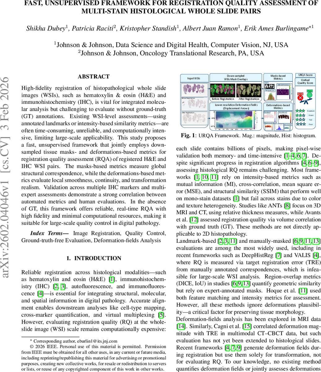
To solve this, the authors propose an unsupervised Registration Quality Assessment framework (URQA) that jointly evaluates (1) global structural alignment using down‑sampled binary tissue masks, and (2) local deformation realism using the deformation field produced by the registration algorithm. The framework consists of two modules:
-
Masks‑based RQA (MRQA).
- Both fixed (H&E) and moving (IHC) WSIs are down‑sampled to the lowest resolution (capped at 512 px) and binarized via Otsu thresholding plus morphological cleaning, yielding masks M_f and M_r.
- Three complementary metrics are computed: Intersection‑over‑Union (IoU), Mean Absolute Error (MAE) between masks, and a histogram‑correlation score (HC) that selects the best of Pearson correlation, histogram overlap, or cosine similarity on grayscale intensity histograms.
- Pre‑defined thresholds (e.g., IoU ≥ 0.80, MAE ≤ 0.07, HC ≥ 0.80) map the three metrics to a discrete score M_Q ∈ {0,1,2,3} (Fail, Poor, Good, Excellent). This provides a stain‑invariant, fast estimate of how well the tissue outlines overlap after registration.
-
Deformations‑based RQA (DRQA).
- The deformation field φ(x)=x+u(x) is extracted from the lowest‑resolution pyramid level.
- Consistency of displacement magnitude M(x) and direction Θ(x) is checked by comparing their standard deviations to the inter‑quartile ranges (IQRs); low variance indicates smooth, plausible motion.
- Jacobian determinant J(x) is computed to assess local volume change: the mean should be near 1, σ_J < 0.25, and the proportion of negative Jacobians (folding) < 1.5 %.
- A smoothness residual (SR) is obtained by subtracting a Gaussian‑smoothed version of the displacement from the original; both the mean and σ of the residual must be smaller than their IQRs.
- Each satisfied criterion contributes one point, yielding a deformation score D_Q ∈ {0,1,2,3}.
The final URQA score Q is defined hierarchically: if either M_Q or D_Q is zero, the pair fails (Q = 0). Otherwise, Q takes the higher of the two scores (1–3). This rule ensures that a registration is accepted only if both global alignment and deformation realism are satisfactory, mirroring how a pathologist would reject a registration that looks globally aligned but contains unrealistic tissue warping.
Experimental validation was performed on an in‑house dataset of 300 H&E–IHC slide pairs covering multiple biomarkers (HER2, cMET, EGFR, PD‑L1). All pairs were registered using the open‑source V‑ALIS algorithm (the top performer in the ACR OBA T 2022 challenge). For quantitative evaluation, 33 slides were independently reviewed by two experts (a board‑certified pathologist and a certified histotechnologist). Experts assigned a binary Pass/Fail label and a four‑level quality grade (0 = Poor, 1 = Fair, 2 = Good, 3 = Excellent).
Key findings include:
- Agreement with experts: URQA achieved the lowest false‑positive rate and the highest true‑negative rate among the three evaluated methods (MRQA alone, DRQA alone, and URQA). In binary classification, URQA’s confusion matrix closely matched expert judgments, especially for the pathologist (E‑1).
- Weighted performance: Using average precision, recall, and F1‑score across the two experts, URQA outperformed the individual modules (e.g., AP = 0.87 vs. 0.84 for MRQA on E‑1).
- Efficiency: Processing times on a standard workstation were 4.3 s for MRQA, 6.3 s for DRQA, and 10.75 s for the combined URQA pipeline on a 512 × 512 down‑sampled slide. Memory consumption stayed below 200 MB, making the method feasible for batch processing of thousands of WSIs.
- Qualitative insights: Visual examples demonstrated cases where MRQA flagged good global overlap but DRQA detected unrealistic folding, leading URQA to correctly reject the pair. Conversely, when both modules agreed, the final score aligned with expert perception of “Excellent” alignment.
The authors discuss limitations: (i) extreme deformations at high magnification (e.g., 20×) may still escape detection because the analysis is performed at low resolution; (ii) Otsu‑based mask generation can be sensitive to low‑contrast or heavily stained backgrounds, potentially affecting IoU and MAE. Future work is suggested to incorporate deep‑learning‑based mask segmentation, multi‑scale deformation analysis, and extension to 3‑D histology or other tissue types.
Conclusion: URQA provides a fast, unsupervised, and interpretable framework for assessing the quality of multi‑stain WSI registration without any ground‑truth data. By jointly measuring global mask overlap and local deformation plausibility, it delivers scores that correlate strongly with expert visual assessment while requiring only seconds of computation per slide. This makes URQA a practical quality‑control tool for large‑scale digital pathology pipelines, enabling reliable downstream analyses such as virtual multiplexing, cell‑type mapping, and quantitative biomarker studies.
Comments & Academic Discussion
Loading comments...
Leave a Comment