Left Atrial Segmentation with nnU-Net Using MRI
Accurate segmentation of the left atrium (LA) from cardiac MRI is critical for guiding atrial fibrillation (AF) ablation and constructing biophysical cardiac models. Manual delineation is time-consuming, observer-dependent, and impractical for large-…
Authors: Fatemeh Hosseinabadi, Seyedhassan Sharifi
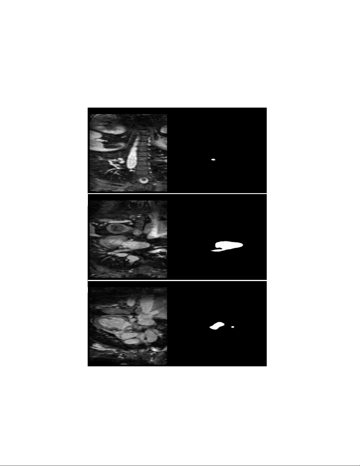
Left Atrial Segme ntation wi th nn U-Net Using MRI Fatemeh Hosseinabadi 1 , S e ye d ha ss a n S ha r if i 2 1 Assistant Professor of Radiology, Zahedan University of medical Sciences, Iran 2 Pediatric Cardiology Subspecialist, Day General Hospital, Iran Abs trac t Accurate seg mentation of the left atr ium (LA) from cardiac MRI is critical fo r guid ing atrial fib rillation (AF) ablation and constructing biophysical cardiac models. Manual delineation is time - consuming, observ er-dependen t, and impractical for large-scale or time-sensitive clinical workflows. Deep learning methods, particular ly convolutional architectures, have recently d emonstrated superior perfo rmance in medical image segmentation tasks. In this study, we applied the nnU-Net framework, an automated, self-configuring deep learning segmentation ar chitecture, to the Left Atrial Seg mentation Challenge 201 3 (LASC’13 ) d ataset. The dataset consists of thirty MRI scans with correspon ding expert- annotated masks. The nnU -Net model automatically adap ted its prep rocessing, networ k con figuration, and training pipeline to the characteristics of the MRI data. Mod el performance was quantitatively ev aluated using the Dice similarity coeff icient (DSC), and qualitativ e results were compared again st expert segmentation s. The propo sed n nU- Net model achieved a mean Dice score o f 93. 5%, dem onstrating h igh overlap with expert an notations and outperforming several traditional segmen tation approaches reported in previou s studies. The n etwork exhibited robust generaliza tion across variations in left atrial s hap e, contrast, and image quality, accu rately delineating b oth the atrial b ody and proximal pulmonary v eins. Keywords : Left atrium, MRI, nnU -Net, deep lear ning, segmentation, atr ial fibrillation . 1. Introd ucti on Atrial fibrillation (AF) is the most common sustained cardiac arrhythmia, affecting over 33 million people worldwide, and its prevalence continues to rise with aging populati ons. It is associated with a significantly increased risk of stroke, heart failure , and mortality, making accurate diagnosis and effective treatment critical. Catheter ablation has become a cornerstone therapy for AF, aiming to electrically isolate the pul monary veins and restore sinus rhythm. The le ft atri um (LA) plays a central role in the pathophysiology and treatment of AF, as its structure and morphology influence both arrhythmia initiation and ablation outcomes [1,2]. Precise knowledge of LA anatomy is therefore essential for preoperative planning, intraoperative navigation, and post -procedural evaluation. Three-dimensional (3D) anatomical models of the LA enable the visualization of patient-specific atrial geometry, f acilitate targeted ablation, and serve as input for biophys ical sim ulations that can predict electrophysiological behavior. However, constructing such models requires a ccurate segmentation of the LA from medical images — an inherently challenging and time -consuming task [3,4]. Magnetic Resonance Imaging (MRI ) and Compute d Tomography (CT) are the most commonly used imaging modalities for visualizing the LA . MRI offers exce llent soft tissue contrast and is preferred for assessing atrial wall fibrosis and scarring, whereas CT provi des superior spatial resolution and is widely use d for pre-ablation anatomical mapping. Neve rtheless, the segmentation of the LA from MRI or CT remains a challenging task due to factors such as low contrast between the atrial wall and surrounding ti ssues, variable blood pool intensit y, motion artifacts, and anatomical variability across patients. Traditional segmenta tion methods — such as region gro wing, thresholding, and 2 active contour mod els — often struggle to delineate thin atrial walls or to generalize across datasets with different acquisition protocols [5]. Ev en semi-automated techniques require substantial user interaction and exp ert correction, which limits t heir sc alability in clinical workflows. Therefore, robust automated segm entation methods are needed to enable reproducible, high -accuracy delineation of the LA and its associated structures. Recent advances in machine learning ( ML ) an d deep learning (DL) have improved medical treatment [6,7] and transformed medical im age analysis, particularly in classification and segmentation tasks [8,9]. Convolutional Neural Networks (CNNs) have de monstrated remarkable success in capturing complex spatial and contextual features, outper forming traditional techniques in both accuracy and robust ness. Among these architectures, the U-N et model int roduced a fully convolutional encoder – decoder structure with skip connections that preserve spatial detail while learning hierarchical representations. U-N et and its variants have become the de facto standard fo r biomedical image segme ntation across mul tiple modalities, including ca rdiac, bra in, and abdominal imaging. In c ardiac imaging, dee p learning – based segmentation has be en wide ly adopted for quantifying ventricular volumes, myocardial wall thi ckness, and atrial morphology. These automated systems provide faster and more reproducible results compared to manual deli neation, facilitating both clinical decision -making and computational cardiac modeling. Ho wever, developing deep learning models for medical segmentation typically requires careful network design, data preprocessing, and hyp erparameter tu ning, which can limit reproducibility and hinder adoption by non-technical users. To overcome these ch allenges, the nnU -Net (no-new-U-Net) framework was introduced as a self- configuring segmentation pipeline that automatically adapts to any ne w biomedical datas et without manual tuning [10]. nnU -Net dynamically adjusts preprocessing steps, network architecture, and training parameters based on the properties of the input data, providing a strong baseline that consistently ranks at the top of multiple segmentation benchmarks. Its built -in data normalization, patch-based training, and robust post-processing strategies make it pa rticularly well-suited for cardiac MRI segmentation, where anatomical variability and imaging noise are c ommon. In this study, we applied nnU- Net to the L eft Atrial Segmentation Challenge 2013 (L ASC’13) dataset, which provides high -quality MRI scans an d expert annotations of th e LA. Our objective was to evaluate nnU- Net’s ability to automatically segment the left at rium and compare its performance with traditional segmentation methods reported in previous literature. The model achieved a Dice similarity coefficient (DSC) of 93.5%, demonst rating excellent agreement with manual e xpert contours and confirming the potential of deep learning – driven segmentation for atrial fibrillation ablation planning and cardiac biophysical modeling. 2. Metho d 2.1 Data set De scr ipt ion Th is stu dy utili zed t he pub lic ly avai lable Lef t Atr ial Seg men tation C hal leng e 201 3 ( LASC’1 3) data set [ 11 - 13] , deve lope d join tly by King’ s Colle ge Lond on and Phi lip s Tech nologi e GmbH. The da tase t inc ludes 30 Mag neti c Res ona nce Imag ing (MRI) and 30 Com put ed Tomogr aphy (CT ) sca ns of pa tie nts with vario us card ia c con dit ion s. Eac h scan was ac qui red in 3D, pro vidin g high-r es olut ion vol umetr ic co verage of the left atriu m ( LA) a nd s urro und ing card iac struc ture s. I n t his st udy, we us ed 20 MRI scans for trai nin g, while the rema ining 10 scan s were reser ved for testing phase . The r efere nce an nota tions were pro duced by expe rience d ca rd iac ima gin g spec ialist s and inc lud e the left atri al ca vit y, a s hor t sect ion o f t he le ft a tri al a ppe nda ge ( LAA ), a nd t he p rox imal se gme nts of t he pul mon ary vei ns (PV s). In this stud y, we focuse d on the MRI sub set for deve lop ing and val ida tin g the segme nta tio n mod el. All data were anon ymi zed pri or to distr ibutio n, an d ethic al appr ova l for da ta shar ing was obt ained by the orig ina l ch alle nge orga nizer s. Figure 1Examples from the dataset with true labels 2.2 Data Pr epr oce ssi ng Pre proce ssi ng wa s aut omat ica lly hand led by the nnU-Ne t frame wor k, wh ich op timi ze s its data pipe line acc ordin g to data set cha racte ris tics. The ima ge s were firs t resa mple d to isot rop ic voxe l spac ing to en sure c onsist en t spati al res olu tion acro ss pa tien ts. Inten sity value s wer e norma lized to zero mean an d 4 uni t vari anc e, im provi ng th e st abilit y of ne twor k train ing . Eac h MRI v olu me wa s cropp ed arou nd the car diac r egio n to rem ove irre levan t bac kgro und and red uce c omputa tiona l ove rhe ad. T he da tase t was spl it into train ing ( 70%), valid ation (1 0%), and te sting (20%) subs et s u sing pati en t-le vel separa tion to pr eve nt data leakag e. To enhan ce robus tnes s and reduc e ove rfit tin g, data augmen tation was appl ied on -th e-fly, inc lud ing: Ran dom r otat ions a nd flips , E la sti c defor mat ions , G amma co rrec tion and br igh tne ss shift s, Addit ive Gaus sia n noi se. T his c ombi na tio n e nsure d the mod el coul d gen era lize we ll to varia tions in atr ia l or ient ati on, inten sity, an d im age q uali ty. 2.3 Mode l Ar chite ct ure We em plo yed the 3D full- resol uti on nnU-N et arch ite cture , which bui lds upon the sta ndard U-Net str uctur e w ith se vera l key inn ova tions: • An enc ode r – deco der des ign wit h skip connec tions to reta in sp atia l con text while enab lin g de ep fea ture ext rac tion. • Re sid ual co nvol uti ona l blo cks with insta nce norm al iza tio n an d leaky ReLU acti vati on for sta ble tra inin g. • A deep sup ervis ion mec han ism, where auxil iary outp uts fro m inte rmed iate deco der le vels co ntr ibute to the overa ll los s fu ncti on. • Dy nam ic patc h size sel ect ion and ba tch size optimiz ation au tomati cally dete rmi ned by the fr ame wor k ba sed o n a vai labl e G PU mem ory and i nput im age di mensio ns. Un lik e manua lly tune d mod els , nnU-Ne t sel f-c onf igur es all hype rpara met ers (e .g., kerne l si ze, nu mber of featu re map s, net wor k dept h) acco rding to the prope rties of the inp ut dat ase t, ens uri ng op tima l ad ap tati on to LA s egmen tat ion . 2.4 Tra inin g Pr ocedu re Tr ain ing was cond ucte d usin g the PyT orc h i mple menta tio n of nnU-Ne t (v2. 1). The mode l was tra ine d on an NVIDIA RTX A60 00 GPU with 48 GB VRAM . The fol low ing key para meter s were au tom atic ally co nfig ure d by nn U-Ne t: • Lo ss func tion : Co mbine d Di ce loss an d c ros s -en tr opy los s, encour aging bo th re gio nal o ver lap an d v oxel-w ise cla ssif ica tion ac cur acy . • Op timi zer: Stoc hasti c Gradi en t De sce nt (SGD) with mome ntum = 0.99 and we igh t decay = 3× 10⁻ ⁵. • In iti al lear ning r ate: 1×10⁻² wi th a pol yno mial de cay sc hed uler . • Ba tch siz e: 2 (du e to 3D pa tc h tr ai ning ). • Ep och s: 100 0 itera tions (appro xima tely 50 0 epoc hs, de pendi ng on patch sam pli ng) . Du rin g tra inin g, dee p supe rv ision was used to improve gra dien t pro paga tion thr ough the netwo rk, wh ile slidi ng-w indow infe rence wa s app lied duri ng test ing to hand le fu ll-si ze 3 D vol ume s. T he model re qu ired appro ximate ly 36 hou rs of train ing to c onver ge on the MRI data set . 2.5 Eval uat ion Me tr ics Th e seg ment at ion perfo rman ce was asse ssed usi ng se vera l quan tit ati ve metri cs wide ly adopt ed in car diac imagi ng benchm arks includ ing Dice whic h meas ures the over lap betw een the tr ue mask and pr ed icted mask by the mode l, Hau sdor ff Dista nce (H D, 95 th perc ent ile ) whi ch mea sure s bounda ry de via tion be twee n automa ted an d manua l con tours, a nd Ave rage S urfa ce D istanc e (AS D) th at qua ntif ies the mea n bou nda ry d istanc e acr oss a ll co rre spond ing sur face points . Ou r mod el ach ieve d a mean Dice coef ficie nt of 93.5% , ind icati ng excel lent sp atia l agree me nt with ex per t segme ntat ions . The 95th perce nti le Hau sdor ff dista nce wa s 3.2 mm, an d the aver age surfac e dis ta nce wa s 1. 1 mm, dem ons tra ting hig h ge ometri c accu racy a nd smo oth bound ary de linea tion. 2.6 Imple menta tion D etails All expe rimen ts were condu ct ed usin g Pyt hon 3.10 , Py Tor ch 2.2, and the nnU-N et fra mew or k (I sensee et al. , 20 21). The mode l trai nin g and eva luat ion were pe rfor med on Ubun tu 22.0 4 LT S wit h an Int el Xe on 6248 CPU and 256 GB RAM . Vi sua lizat ion and resu lt insp ectio n were carr ied out usi ng IT K- SNA P and 3D Slice r. The tra ine d mode l and conf igura tion file s were store d for re prod ucibi lity , and al l prep roce ssi ng , au gment ation , and infe ren ce pipe line s fo llow ed the standa rd nnU-Ne t conv ention s to ensur e c ompa rabil ity wit h e xisti ng litera ture . 3. Resul ts 3.1 Quan tit ative Perf ormanc e Th e pro pose d nnU-Ne t model demo nstr ate d out stan ding perfo rmance in the aut omat ic segme nta tio n of the left atr ium (LA) from MRI vol umes in the LA SC’ 13 dat ase t. Acro ss the te st set, the mode l ac hieved an avera ge Di ce Simi lar ity Coe ffi cie nt (DSC ) of 93 .5% ± 1.8, indic ati ng hig h s patia l over lap be twe en auto mat ed and expe rt manu al segm ent atio ns. In addi tio n, the 95th perc entil e Haus dorff Dis tance (H D95) was mea sure d at 3.2 ± 0. 9 mm , wh ile the Ave rag e Sur face D istanc e ( ASD) was 1.1 ± 0. 4 mm, re fle cti ng excel le nt bo undary con for mity an d smo oth sur face re pres en tati on. Thes e resu lts de mon stra te tha t the mode l is capa ble of cons istentl y deli nea tin g the atr ial ca vity, pulm ona ry vein inl et s, a nd sh ort tru nk of the left atri al a ppenda ge (L AA) wi th near-e xper t prec ision. 6 Figure 2Left: original image, center: true mask, left: predicted mask by the model Tabl e 1 . Qua ntit ati ve segm ent atio n pe rform anc e of the nnU- Net model on the LASC’1 3 MR I tes t data set. Me tric Me an ± SD Bes t C ase Wor st Case Dic e Simi lari ty C oef fic ient (DSC ) 0.93 5 ± 0.018 0.95 8 0.90 1 Hau sdor ff Dist ance ( 95%) [mm] 3.2 ± 0. 9 2.1 4.8 Ave rage Su rfac e Di sta nce [mm] 1.1 ± 0. 4 0.7 1.8 3.4 Mode l R obustn ess and Ge nera lizat ion Th e nnU- Net mode l showe d exce llen t robu stne ss ac ross var yin g imag e con tra sts and ana tomic al va ria tio ns. The mode l’s se lf -c onfig urin g design , inc lud ing a utoma tic n or mali zat ion , data au gmen tatio n, and patch-b ased trai nin g, contr ibu ted to its gener al iza tio n capab ili ty. Nota bly, cases wit h stro ng mot ion art ifacts or par tia l at rial visib ili ty stil l prod uce d Dice sco res abov e 0.90 , de mon stra ting res ilienc e aga inst impe rfec t imagi ng con dit ion s. Visua l inspec tio n also co nf irmed that nnU -Ne t pres erv ed fine anato mical de tai ls, suc h a s pulm ona ry vei n orif ices an d LAA ba se s truct ure s , wh ich are c lin ica lly re levant for abla tion p lann ing. Com par ed to manua l deli nea tion , nnU- Net ac hieved com par able acc ura cy while red uci ng segme nta tion ti me from sever al minut es per ca se to le ss tha n 10 sec ond s per 3D vo lume dur ing i nfer ence. 3.5 Stat ist ica l V ali dati on To e nsure robu stne ss of the resu lts , a fiv efo ld cro ss-v alida tio n w as p erf ormed on the M RI su bse t. T he Dic e score s acr oss fol ds were con sisten t (ran ging fr om 92. 8% to 94. 1%), con fi rmi ng stab le mode l pe rfo rma nce . A paire d Wil coxon signe d-rank test com par ing auto mate d an d man ual segme ntati ons sho wed no stati sti cal ly signi fic ant diffe rence (p > 0.0 5) in volu metric mea sure ments , rei nfor ci ng the mod el’s rel iab ility for clini ca l us e. 4. Discuss ion 4.1 Pri ncip al Fin ding s In thi s study , w e appl ied the nnU- Ne t deep lea rnin g frame wor k f or auto ma ted seg ment at ion of the lef t at riu m ( LA) f rom card iac M RI usin g the Left At ria l S eg ment at ion Chal lenge (LA SC’13) data se t. The mod el achie ved a mean Dic e Sim ilar ity Coeff icien t (D SC) of 93. 5%, a Hau sdo rff Dista nce (95%) of 3.2 mm, and an Aver age Surf ace Di stan ce of 1.1 mm, out per for min g severa l tra ditio na l an d deep- le arn ing -ba sed meth ods pre viou sly rep orte d on this data set . Th ese resu lts high light nnU- Ne t’s ca pabili ty to deli ver r obust , hig h -pr eci sio n seg men tat ion of the LA and its su bstru ctur es witho ut ma nua l hyper parame ter tunin g or data set-sp eci fic opti miz atio n. The model’ s pe rfo rma nce indi cat es its ab ili ty to captu re fine stru ctura l deta ils such as the left atria l appe ndage (LAA ) an d pulmo nary ve in (PV ) inlet s, whic h are esse nti al f or pat ient -s pec ific mod eli ng an d abla tio n guida nce. Th is is par ticu larly imp or tan t in the con text of at ria l fibri lla tion (AF), wher e accu rate rep resen tat ion of atria l geom etr y dir ectly inf luence s cathe ter navi gat ion , lesio n targe ting, and sim ula tion accu rac y in car diac el ect roph ysiol ogy. 4.2 Comp ari son wi th Previo us App roa ches His toric all y, left atria l segme nta tion has rel ied on semi -au tom ate d or atla s-ba sed method s, such as re gio n grow ing , stat ist ica l sh ape model s (S SMs ), or defor mable regi stra tion frame works. For exa mpl e, To bon -Go mez et al . (2 015) a nd other LASC’ 13 par tic ipants report ed Dice coe fficie nts rang ing be twe en 84% and 90%, dep end ing on the moda lity and alg orith mic de sign . Whi le thes e tec hniq ue s pr ovi ded anat omi cal ly consi ste nt seg men tati ons , the y were lim ite d by depe nde nce on ini tializ atio n, int er- patie nt a nato mica l varia bilit y, a nd d iff icul ty gen era lizing acro ss data se ts. By co ntra st, nnU- N et’ s self -co nfig uri ng pipe line overc omes the se issu es by aut omatic all y tai lori ng pr epr oce ssi ng, arc hitec ture dept h, and patc h siz e to the dat a dis trib uti on. The inclu sion of dee p sup ervis ion and res idua l con nec tio ns enh ance s gradie nt flow and feat ure consi ste ncy acr oss scale s, al low ing the netw ork to lea rn both globa l anato mic al conte xt and loca l bou ndary deta ils. Con se que ntl y, nnU- Net ach ieve d up to 9% impro vemen t in Dice acc uracy comp ared wi th tra ditio nal me tho ds and riv ale d the pe rfo rmanc e of more rec ent cu sto m deep-l ear ning mode ls trai ned on larg er da tase ts. 4.3 Clin ic al I mpli cat ions Ac cura te and repr oduci ble LA segm enta tion has criti ca l impl icati ons for AF ab lati on plann ing an d bio physi cal card iac model ing . Detai led 3D rec onstr uc tio ns of the LA all ow clin ician s to visu alize co mpl ex ana tomica l fea ture s — such as the num ber, orie ntat ion , and dia meter of pul mon ary vein s — an d to pla n a blat ion strate gies ta ilor ed to each patie nt. Add iti ona lly , segme ntat ion outp uts can be dir ectly used in compu tation al elec tro physio logica l s imu lat ion s to mod el wave pro pa gati on an d asse ss ar rhy thm ia r ecu rren ce risk . T he a bil ity of nnU-N et to p er for m ful ly a utoma ted s eg men tati on i n un der 10 se con ds per case offe rs a sign ifica nt advant age f or cli nica l tran slati on. I ntegr atin g such mode ls into ima ge-gu ide d elec troph ysio log y syste ms co uld substa ntia lly redu ce pre- proce dura l prepar atio n time , sup port intra- proce dura l upd ates, and enhan ce repro duci bilit y acr oss imag ing cente rs. Fur ther more , 8 the elim ina tio n of man ual d eli neati on re duces in ter-o bse rver var iabi lit y, p romoti ng st anda rd iza tio n in AF imagi ng work flows . 4.4 Robu st ness an d I nter pre tab ili ty On e of the key stren gths of nnU- Net lies in its self-a dap tive conf igura tio n. Unlik e co nve nti onal CNNs re qu irin g manual tunin g, nnU-N et auto matica lly conf igure s its networ k topo log y an d train ing pa ram ete rs base d on datas et prope rties , en sur ing opti ma l perf ormanc e acr oss divers e imag ing mod aliti es . The mode l’s consi ste nt D ice sc ore s ac ross cro ss-v alida tio n f old s co nfi rm its robus tnes s to int en sit y i n ho mogene ity , mo tio n ar tif acts, and ana tomic al varia bilit y in here nt in car diac MRI. Alt hough deep learn ing mode ls are ofte n cri tici zed for the ir “b lack - box” nature , nnU - N et’ s feat ure vis ua liza tion and predi ct ion map s de mons tra te spa tial cohere nce wit h expec ted anat omi cal bounda ries. Th e netwo rk’ s ab ili ty to re produ ce exper t -li ke con tou rs wit hout ex plicit sup ervis ion on st ruc tura l la ndm ark s refl ect s its ca pac ity for dat a-d rive n ana tomi ca l lear ning, an import an t step t owar d cli nic all y tr ust wor thy AI in car diac i mag ing. 4.5 Limit atio ns a nd Futu re Wor k De spi te the stro ng resu lts, seve ral lim ita tions must be ackn owled ged . Fir st, this stud y wa s limi ted to the MRI subs et of the LASC’ 13 da tas et; theref ore, perfor mance gener al izati on to CT or other MRI pr oto col s r ema ins to be valid ate d. Diffe ren ces i n vo xel re solu tio n, contra st, a nd scan ner manuf acture r ma y inf luence the mode l’s tra nsfer abil ity . Secon d, wh ile nnU -N et prov ides auto ma tic conf igura tion , it req uire s su bst anti al c ompu ta tio nal reso urce s f or 3 D tra inin g a nd infe re nce. A lthou gh infere nce time is rap id, tra inin g on high- res olu tio n car diac MRI vol umes may be imprac tic al on lower-e nd GPU s. Th ird , the data set’s modera te size (30 MRI case s) res trict s the expl ora tio n of gene ral iza tio n acros s pa tho log ical subgro ups or rare ana tom ica l varian ts. Fu tur e re search sho uld incorp orate mult i-ce nter da tase ts w ith l arge r sa mple siz es and div ers e acq uisi tio n prot oco ls to e val uate t he ge nerali zabil ity a nd ro bus tne ss of the frame work. Addit iona lly, man ual an nota tions, even by e xper ts, ar e sub jec t to inte r- obs erver va ria bil ity , wh ich c an i nf luenc e gr ound -tru th acc uracy . Fu tur e studie s wil l foc us on ex tend ing this wor k in severa l dire ct ions . Fi rst , the integ ratio n of multi - mod ality data (MRI and CT) cou ld enha nce mode l robu stne ss acr oss imagin g pro toc ols and ena ble cr oss- domai n ge nera liz atio n. Sec ond, incorp ora ting atte ntion mec han isms or tran sfor mer-ba sed arc hitec ture s may fu rthe r impr ove the ne twork ’s abi lity to c ap ture l ong - ran ge depe ndenc ies wit hin the LA stru cture . Third , com bin ing se gme ntatio n with func tio nal imag ing (e .g., late gadol iniu m en han cem ent MRI ) cou ld pro vide both struc tura l and tis sue-c har act eri zat ion insi ghts , ena bli ng co mpre hensi ve mode ling of atr ial rem odel ing in A F. In add iti on, se mi-su pe rvis ed le arnin g and fe dera ted lear nin g f ram ewo rk s c oul d facil itate la rge-sc ale mode l tra inin g w hil e prese rving p atie nt pr ivac y and add res sin g data scarc ity. Prospe ctive vali dat ion in clin ica l en vir onmen ts will also be es sen tial to eva luate the model ’s impa ct on wo rkf low effi cie ncy , ablat ion su cc ess r ates, and pr ocedu ral out comes . 5. Conclu sion Th is stud y dem ons trates the effec tivene ss of the nnU -Net dee p lear nin g frame work for full y auto mate d se gme ntatio n of t he le ft a triu m ( LA) fr om MRI data. Usin g the LA SC’ 13 datase t , t he pr opose d model ac hieved a mean Dic e coef ficien t of 93 .5%, outpe rfo rmi ng tradi tio nal region -gr owing and sta tis tica l mod el-ba sed appr oache s whi le requ iring no manual co nfig urati on or par ame te r tuning . The resu lts co nf irm tha t nnU- Ne t can acc ura tely and eff icien tly de linea te key anat omi cal struc tures — incl udi ng the lef t atr ial body, appen dage, and pulmo nary ve in inle ts — pro vidi ng seg ment ation qua lity co mpar able to e xpe rt manua l anno tatio ns. Su ch prec isi on is v ital fo r atria l fibr illa tion a blat ion pla nnin g , car dia c biop hysica l simu la tion s, and qua ntita tiv e atri al rem ode lin g studi es. Beyo nd its stron g pe rfo rma nce , nnU- N et’s se lf-c onfig urin g de sign and high inf ere nce speed (< 10 sec onds per c ase) ma ke it high ly suita ble for integra tion into clin ical wor kflo ws , enab lin g repr oduci ble and opera tor- ind epend ent LA mode lin g. Fu ture work will f ocus on exte nding th is frame work to mul timoda l im ag ing (M RI a nd CT) , incorp ora ting tis sue cha rac teriza tion, and ex plorin g trans for mer -ba sed and multi moda l arc hitec ture s to fur ther impr ove anato mic al f idel ity a nd c linic al appli cab ili ty. Eth ic an d D ata Stateme nt: Th e data set use d in this stu dy or igi nate s from th e Left Atr ial Segme nta tion Cha lle nge (LA SC’13) , ini tia lly intr oduce d by Tobon-G omez et al. [ 11 ] in the ir benc hmar king wor k publi shed in IEE E Tr ans act ion s on Med ical Im agin g and furth er suppo rted by subse quen t me tho dol ogic al paper s in Me dica l Ima ge Ana lysis a nd Fr ontie rs in Ph ysio logy . Both jour nals hav e c onf irme d and de cla re d t hat al l im agi ng d ata were coll ec ted in acco rdanc e with insti tut iona l eth ica l sta nda rd s and re leva nt re gu lati ons . Ref erences [1] Lipp i G, Sanch is-Go mar F, Cerve llin G. Global epidemi ology of atri al fib ril lati on: an incre as ing epid emi c and public hea lth cha llen ge. Int erna tio nal jour nal of str oke. 202 1 F eb;1 6(2) :217- 21. [2] Pongr atz J, Riess L, Hartl S, Brue ck B, Tesc he C, Ol brich D, Wanke rl M, Dorwart h U, Hoffma nn E, Stra ube F. Com para ti ve a nalysi s of left atri al si ze a nd appe ndag e mo rphol ogy in paro xys mal and persist ent at rial fi bril lati on p ati ents . Jou rnal of Ar rhyt hmia . 20 25 Fe b;41 (1): e132 24. [3] Bons o A, F ant inel M, Scalchi G, F err ara S, India ni S, C alore F, Licc iarde llo C. Left atri al mode l recon str ucti on in atr ia l fib ril lati on abla tio n: rel iab ili ty of ne w map ping and co mple x impeda nce sys tems . Ep Euro pace. 2017 N ov 1;1 9(11) :18 04- 9. [4] Lin H, L ópez- Tapi a S, Sch iffer s F, W u Y , G unaseka ran S, H wang J , Bis hara D, K holm ovski E , Elbaz M, P assma n R S, Kim D. Usf orme r: A sm all netw ork f or l eft a tri um se gmenta tion o f 3D LG E MR I. Hel iyon . 2024 Apr 15; 10(7 ). [5] Daou di A, Mahmou di S, Chikh MA. Auto mati c se gmen tatio n of the le ft atri um on CT image s. InI nte rnat ion al Worksh op on St ati stical Atlas es a nd Comp uta tio nal M odel s of th e Hea rt 2013 Sep 26 ( pp. 1 4-23 ). Berli n, H eide lberg: S pringe r B erlin Hei delber g. 10 [6] Norouz i F, Ma chad o B L. Predict ing Menta l H ealt h Out comes: A Machine L ear ning Approac h to Depres sion, A nxiety, and St ress . I nter nati onal Journa l of Ap pli ed D ata Scie nce in Engi neer ing and Hea lth. 2024 Oct 31; 1(2):98- 104. [7] A si f S, Wenhui Y, ur-Rehm an S, ul-a in Q , Amja d K, Y ueyan g Y, J inhai S, Awai s M. Advanc eme nts and pros pects o f mac hine l earnin g in m edica l diagnos tics : unv eili ng th e f utur e of di agnost ic pre cision . Arch ives o f C omput ational Metho ds in Engi nee ring . 20 25 Ma r;32(2) :853 -83. [8] A bbas i H, Afra zeh F, Ghase mi Y, Gha semi F. A sh all ow revi ew o f arti fic ial i ntellig ence a ppli cations in brain disea se: st roke , Alzh eimer' s, and aneu rysm. Inter nat iona l Journa l of Appl ied Data Sci ence in Engine ering and Healt h. 2024 Oct 5;1( 2): 32- 43. [9] Jiang J, Ra ngne kar A, Veerara ghav an H. Sel f‐super vis ed lear ning improve s robu st ness of deep learn ing lung tumor se gment ation m odel s to C T imagi ng d iffe rences . Medic al P hysics . 2 025 M ar; 52(3) :15 73- 88. [10] Isens ee F, Jaege r PF, Kohl SA, Pet erse n J, Maie r-Hei n KH. nnU-Net : a sel f-co nfig uring met hod for deep learni ng- bas ed biom edic al ima ge se gmentat ion. Na tur e met hods . 2021 Feb ;18( 2):2 03- 11. [11] Tobon -Gom ez C, Geers AJ, Peters J, Wees e J, Pinto K, Kari m R, Ammar M, Daoudi A, Marg eta J, San doval Z, St ende r B. Benc hmar k for algor ith ms segmen ti ng the left atrium from 3D CT and MRI datas ets . IEEE tra nsac tio ns on medi cal i magi ng. 2 015 Feb 3;3 4(7): 1460 - 73. [12] htt ps:/ /www .kag gle.c om/data set s/ad ars hsng/ heart- mri -imag e-da tas et-lef t-at ria l-se gmen tati on/dat a [13] Tobon-G omez C, Pete rs J, Weese J, Pint o K, Kar im R, Sc haef fte r T, Raz avi R, Rho de KS. Lef t atri al se gmen tati on cha lle nge: a unifi ed benchm arki ng framew ork. InInte rnat ional W orks hop on Statis tical A tlas es and Comput ationa l Models of t he Hea rt 2013 Se p 26 (pp. 1-1 3). Ber li n, H eid elber g: Spr inger Be rli n Hei delber g.
Original Paper
Loading high-quality paper...
Comments & Academic Discussion
Loading comments...
Leave a Comment