Simultaneous use of Individual and Joint Regularization Terms in Compressive Sensing: Joint Reconstruction of Multi-Channel Multi-Contrast MRI Acquisitions
Multi-contrast images are commonly acquired together to maximize complementary diagnostic information, albeit at the expense of longer scan times. A time-efficient strategy to acquire high-quality multi-contrast images is to accelerate individual seq…
Authors: Emre Kopanoglu (1, 2), Alper G"ung"or (2
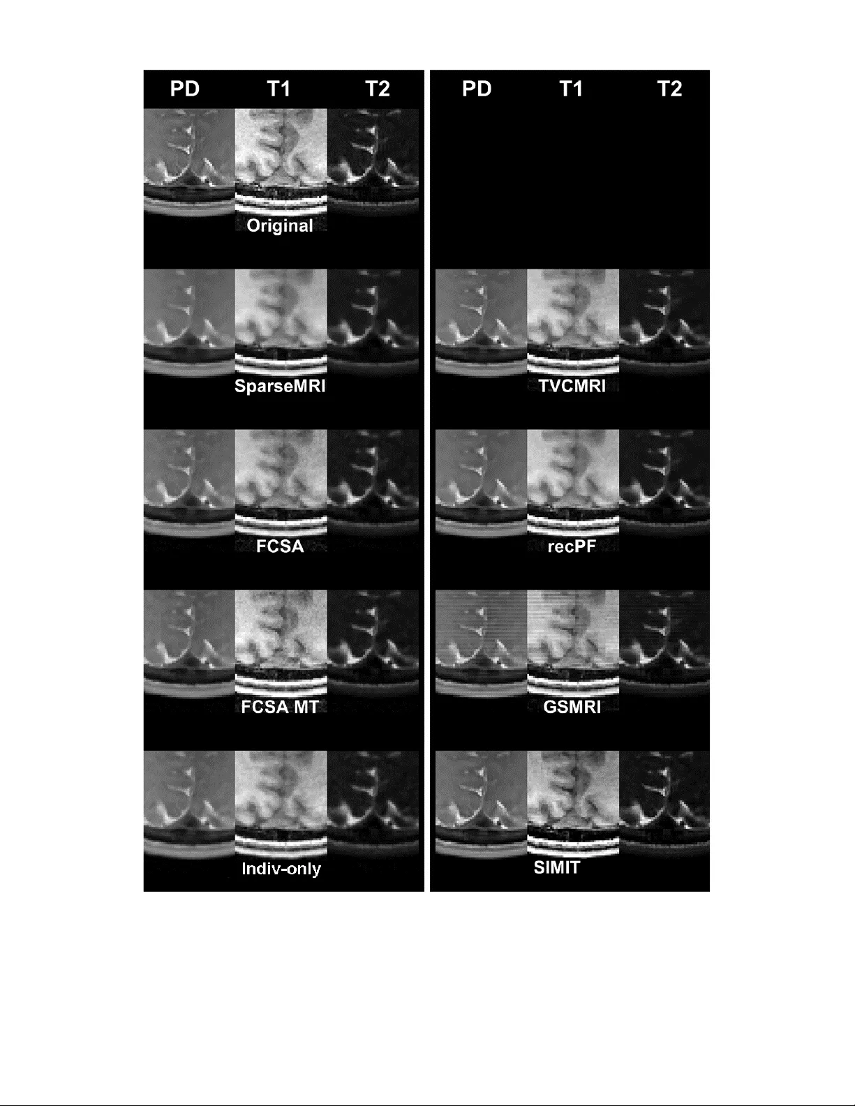
1 1 Simultaneous use of Individual and Joint Regularization Terms in Compressive Sensing: Joint Reconstruction of Multi-Channel Multi-Contrast MRI Acquisitions Emre Kopanog lu 1,2,* , Alper Güngör 2,3 , Toygan Kilic 3,4 , Emine Ulku Saritas 3,4,5 , Kader K. Oguz 4,6 , Tolga Çukur 3,4,5, § , H. Emre Güven 2, § 1 Cardiff Univ ersity Brain Research Im aging Centre (CUBRIC), Schoo l of Psychology, Cardif f University, Cardiff, UK 2 ASELSAN Research Center, Ankar a, Turkey 3 Departmen t of Electrical and Electron ics En gineering, Bilkent Un iversity, Ankara, Turk ey 4 National Magn etic Resonance Research Center (UMRAM), Bilken t University, Ank ara, Turkey 5 Neuroscience Program, Sabuncu Brain Research Center, Bilkent University, An kara, Turkey 6 Departmen t of Radiology, Hacettepe Univer sity, Ankara, Turkey Running Hea d : SIMIT: Joint Reconstruction of Multi- Channel Multi- Contrast MRI Images * : To whom co rrespondence should be addressed. Corresponding Autho r: Emre Ko panoglu Address: CUBRI C, Cardiff Univer sity, Maindy Road, Card iff, CF24 4H Q, UK e-mail: emre.kopanoglu@gmail.com § : These senio r authors co -supervised this study. Words: 226 (abstract), 5969 (body) Figures: 12 2 2 ABSTRACT Multi-contrast images ar e com monly acquired tog ether to maximize co mplementary diagnostic information, albeit at the expense of longer scan times. A time- efficient strategy to acquire high- quality multi- co ntrast images is to accelerate individual sequences and then reconstruct undersampled data with joint regularizatio n terms that leverage common information ac ross con trasts. Ho wever, these term s can cause fea tures th at are unique to a sub set of co ntrasts t o leak in to the o ther contrasts . Such leakage- of -features may appear as artific ial tis sues, th ereby misleading d iagnosis. Th e g oal o f this study is to dev elop a compressive sensing method for m ulti- channel multi-contrast magnetic resonance imaging (MRI) that optimally utilizes shared information while preventing feature leakage. Joint regularization terms group sparsity and colour total variation ar e used to ex ploit common features across images while individual sparsity and total variation are also used to prevent leakage of distinct features across contrasts. The multi-ch annel multi-contrast reconstruction problem is solv ed via a fast algorithm based on Altern ating Direction Method o f Multipliers. The p roposed method is compared against using only individual and only joint regularization terms in reconstruction. Comparisons were performed on single -channel simulated an d multi-channel in -vivo datasets in terms of r econstruction quality and neuroradiolo gist reader scores. The proposed method demonstra tes rapid convergence and improved image quality fo r both simulated and in -vivo datasets. Furth ermore, while reconstructio ns that solely use joint regularization term s are prone to leakage- of -f eatures, the proposed method reliably avoids leakag e via simult aneous use of joint and ind ividual terms, thereby holding great promise for clinical use. Index Terms: compressive sensing , joint reconstruction, leakage - of -features, magnetic resonance imaging, multi co ntrast, parallel imag ing, image reconstruction , joint regularization . INTRODUCTION Multiple imag es of the same anatomy under the influence of different con trasts are of ten acquired in magn etic resonance imaging (MRI) to accumulate d iagnostic informatio n. Common ex amples includ e multi -contrast imaging with T 1, T2, or PD weighting, param etric m apping, and d iffusion weighted imaging. Howev er, with each acquisition lasting several minutes, MRI exams can become impractical ly long and costly. Pro longed scan times also increase suscept ibility to patien t motion and necessitate cumbersom e motion co rrection or image registration procedures. T herefore, scan -time reduction techniqu es are d irely needed to limit cost, patien t discomfo rt, and motion with increasing n umber of acquisitions. 3 3 Accelerated imagin g approaches including p arallel imaging (PI) 1-6 , multi-slice imagin g 7-9 localizing the excitat ion volume 10- 17 , dep hasing outer volumes 18,19 , localizin g encoding to a sub -volum e 20-22 and compressive sensing (CS) 23-45 are promising solutions. Among these, CS h as gained prominence in the last decade, as it do es not require complicated excitation pulses with increased specific ab sorption rate (SAR) or addition al hardware. Furthermore, CS is com patible with alternative approaches such as parallel imagin g 32,41 and simultaneous multi -slice imaging 46 . Convention al CS techniq ues process each ac quisition in a m ulti-co ntrast pro tocol individually 25-28 . Yet, altho ugh tissues may appear at d ifferent signal levels in separ ate contrasts, th e underlyin g tissue stru cture is shared am ong multi-co ntrast acquisitions. As such, multi-co ntrast images share co mmon tissue boundaries, and they are likely compressible in similar tran sform domain s. These observations have motivated resear chers to investigate th e benefits of jo intly reconstructing multiple imag es of the same anatomy 29-40 . Pro posed applicatio n domain s include dynamic MRI rec onstructions that handle single-contrast acquisitions across time 35,36 , multi-coil MRI reconstructions 32 , multi-echo MRI reconstruction s that h andle repeated acquisitions with minor changes in contr ast 33,37 , fat- water separation 38 , m ulti-contrast MRI reconstruction s that pr ocess multiple distinct con trasts 29-31 , and even multi-mod ality reconstruc tions 34 . Joint recon structions aim to utilize th e inform ation shared across contr asts to improv e image quality. Sp arse recov ery during joint recon structions has been attempted with a multitude of reg ularization t erms in literature . A group of studies h ave focused on aggregat ion of individual regularization terms o n each separate image such as the well -known -sparsity 47 and Total Var iation (TV) 48 terms . In Ref. 32 , sparsity was promoted simultan eously ac ross multiple receive channels b y imposing -sparsity on concatenated multi-channel dynamic MRI data. Ref. 38 j ointly reconstructed water and fat images from a multi-echo acquisition by minimizing the sum of individual regu larization function s on each imag e. Ref. 49 perfor med a quasi- joint recon struction by spatially weighing the individual -sparsity and Total Variation o f an image using structural information ex tracted from a prior individually reconstructed imag e. The performance im provement with joint reconstructio n depends on how shared inform ation is leveraged against the information unique to each contrast. Classical individual regularization terms help preserve unique information in each contrast without leakag e of distinct features ac ross images, but reconstructio n s ca n be sub -optimally sensitive to shared information across contrasts. Mean while, joint reg ularization terms such as gr oup sparsity 50 and Colour TV 51 that enforce -sparsity and total variation on multiple images simultaneously have provided useful in several applications including parametric mapping 40 , diffusion tensor imaging 39 , mu lti-echo T2-weigh ted imaging 29,33 and mu lti-contrast imaging 30,31,52,53 . Ref . 35 minimized the nuclear norm to exploit the temporal correlations whereas Ref. 36 used Frobenius and nuclear norms in a blind compressed sensing approach to dynamic MRI. Variations of TV reg ularization such as total gene ralized variation 34 , parallel sets 54 and weighted joint (colour) TV 55,56 h ave also been used for join tly r econ structing MR and PET images 34,54,56 o r f or multi-co ntrast MRI 57,58 . Joint regularization terms boost sensitivity for featur es that are common across acquisitions, but as a result th ey can reduce sensitivity 4 4 for features t hat are unique to each acquisition, and a feature that is on ly pro minent i n a single acquisition may l eak into reconstruction s of o ther acquisitions. Appearance of such artificial features can sev erely im pair diagnostic evaluations; therefor e, multi-acqu isition reconstructions sho uld be carefully inv estigated for leakage - of -features. In this study, we propo se a reconstructio n method for multi -acquisition MRI, named SIMultaneous u se o f Individual an d joinT regularizatio n terms for joint CS - PI reconstruction ( SIMIT). SIMIT leverag es both join t and individual regularization terms to maximize sensitivity for shared f eatures among co ntrasts as well as unique features of each contrast while p reventing undesirab le leakage- of -featu res. Specifically, colour TV ( CTV) 51 an d gro up -sparsity 50 ar e used to explo it co mmon information acr oss contrasts, and ind ividual TV 48 and -sparsity 47 are used to prevent leakage- of -features. SIMIT is demonstrated for mu lti-contrast imaging, where the resu lting optimization probl em is so lved efficiently via an adap tation 59 of Alter nating Dir ection Method of Multipliers ( ADMM) 27,60,61 . First, SIM IT is compar ed against alternative reconstructions that only use indiv idual -sparsity and TV terms (Indiv -only) 59 or only use th e joint terms CTV and group -sparsity ( Joint-only) 62 , on a numerical p hantom dataset . The phantom only includ ed a single -chan nel receiver coil to isolate po tential leak age artefacts . SIM IT is th en compared again st Indiv-on ly and Joint-only as well a s ESPIRiT reco nstructions 63 on multi- channel in vivo datasets. The main contribution s of this study are as f ollows: 1) We i ntrodu ce the simultaneo us use of individual and join t versions of regularization terms in a multi- channel multi-acquisition reconstruction problem. 2) We demonstrate improved image quality and r eliability against leakage- of - features in acceler ated mu lti- co ntr ast MRI . Sing le-channel and multi-chann el implemen tations of this method were presented in part in the 2017 and 2019 Annual Meetings of the Intern ational Society fo r Magnetic Resonan ce in Medicine 64,65 . In Ref. 64 , a single-chan nel version of the metho d was pr esented on a single subject. In Ref . 65 , preliminary comparisons were performed between an earlier multi -channel implementation and ESPIRiT. Here, further to Refs. 64,65 , we p rovide a d etailed theoretical description of the multi-channel, multi-co ntrast reconstruction method, thoroughly investigate the benefits of simultaneously using individual and joint regularization terms as opposed to u sing only joint or o nly in dividual terms, dem onstrate r econstruction performance across a broad range of acceleration rates and numbers of jointly reconstructed contrasts, and perform q ualitativ e and quantitative co mparisons ag ainst three state- of -the- art methods for in-v ivo data acquired from N=11 p articipants. METHODS Theory We pro pose to jointly reconstru ct multi -contrast datasets by leveragin g common information acro ss contrasts via CTV and group sparsity ( -norm , denoted by ) regularization while preventing unwan ted leakag e artefacts v ia indiv idual TV ( and sparsity ( -norm, denoted by ) regular ization. The resulting optimization problem is: 5 5 (1) , (2) where is the num ber of contrasts, is the number of c hannels (coils), , , and denote th e encoding matrix, the reconstructed image vector , the received signal acquir ed through channel fo r contrast and the up per-bound for data-fidelity. Equation (2) d enotes the data fidelity constraint fo r the th -contrast an d -channel. We prefer including data -fidelity as a constraint as opposed to a La grangian form, since can simply be set accordin g to the noise level ca lculated fr om noise-only data (i.e., dat a acquired with out RF excitation). Th e CTV, , TV, and regularizatio n terms can be ex pressed as: , , , , (3) where denote the respective regu larization parameter s. In (3) , denote the image g radients in two orthogonal d imensions. Note that, all f unctions in ( 3) can trivially b e extended to a h igher number of dimensions a s wo uld be required for three-dimen sional or dynamic acquisitions. The jo int regularization terms an d CTV combine the c ontrasts or their spatial derivatives, respectively, before reg ularizing the images. This allows an image wit h an incon spicuous tissue boundar y that would normally b e suppressed via TV regularization to retain this boundary, if the boundary is mo re prominen t in anoth er co ntrast . However, a p rominent but unique fea ture in one contrast may lead t o imperf ect no ise suppression in other contrasts, leading to misleading artificial tissues . I.e. , joint r egularization terms enhance reconstruction s based o n common properties across co ntrasts but may also lead to leakage o f unique f eatures across contrasts. Meanwhile, the indiv idual regularization terms TV and supp ress interference and noise based on the uniqu e structur al prop erties of each co n trast, pr omising sparse rec overy at higher undersamp ling rates withou t introdu cing unwan ted feature transfer ac r oss contrasts. Therefo re, the simultaneous use of the individual terms with the joint terms suppress es leak age- of -features due to the latter. Note that, while the individual and grou p sparsity terms can be applied in a transform domain that sparsifies the image, they are applied in the image domain in SIM IT, since empirical results in the development stages showed > 3 dB b etter peak signal- to -noise ratio (pSNR) when these terms were applied in the image d omain rather than in Wavelet or Discrete Cosin e Transfo rm domains. Note th at, for typical Wavelet transforms in 6 6 MRI reconstru ction, the -term in Wavelet domain captures partly similar informat ion to the TV ter m in image domain . This redundancy is further increased due to simultaneous use of individual and joint versions of regularization terms . In contrast , an - term in the image d omain can impro ve capture of uniqu e prior inf ormation ov er t he TV term. In this study, p SNR was ca lculated as the ratio of the maximu m image intensity acr oss vox els to the root- mean-squared error b etween th e original ( ) an d the reconstructed ( ) images , and expressed in decibels : . In the Supporting Information, we give the general ADMM formulation, show how the proposed optim ization problem for mu lti - contrast MRI can be cast in this general formulation, and derive the u pdate ru les for im plementation. The onl y pa rameter not shown in the bod y of the manuscript, , is the step- size parameter which determines the rate of convergen ce; a smaller µ m eans larger steps and faster convergence. ADMM is known to c onverge under mild conditions 66 and t he step size should be carefully selected to ensure good convergence behaviour, as the algorithm may diverge for ver y small µ . An automated way o f selecting this parameter is given in 67 . For non-convex problems, if the exact solutio n of each sub-problem is kn own, then the algorithm converges to a local min imum. Undersampling Mask s and Noise k-Space data wer e retrospectively un dersampled in one ( 1D-acceleration) or two (2D- ac celeration) phase- encode directions to demonstrate perform ance for two- and three-dimensio nal imaging, respectively (Fig. 1). The central one- eighth section of k-space was fully-samp led. Fo r 2D -acceleration, the diameter of th e fully-sampled disc was set to one-eig hth of the width of th e k -space. Sampling masks were generated using probability distribution fun ctions that decaye d with a po lynomial order of (R-2) or 3 (whichever is larger) , where is th e undersamp ling factor 25 . The und ersampling masks were id entical across reco nstr uction methods bu t were different across co ntrasts . Fig. 1. Two examples of 1D and 2 D-undersamp ling masks. The data-fidelity u pper-bounds for SIMIT ( ) an d its alternative variants (individual terms: Indiv-only; joint ter ms: Joint-only) were empirically set to half of the square root o f the n oise power in experimental reconstructions. Note that simulations were designed to investigate dif ferent factors that may aff ect performance. To isolate the effect of such factors on reconstruction 7 7 performan ce, noiseless images wer e used for the simulations , unless specified other wise. Numerical phanto m The num erical dataset was gen erated using a realistic individual - subject brain phantom that contained segmentation masks for eleven types of tissues 68 . Th e o riginal data for the phantom were acquired using a 1.5T scanner with th e fo llowing p arameters: T1 -weighted images ( 3D sp oiled grad ient ec ho ): TR, 22ms; T E, 9 .2ms; flip-an gle, ; reso lution, 1mm isotropic ; PD - and T2- weighted images (turbo spin echo ): TR, 3300ms; TE ( PD/T2), 15 /104ms; resolution, 1mm iso tropic; number of slices, 62; slice thickness, 2mm ; interslice gap, 0mm . The following contrasts were simulated : PD -weig hted (TE/TR: 17/27 75 ms), T1 - weighted (TE/TR: 14/575 ms), T2 -weighted (TE/TR: 102/2775 ms), T1-weighted fluid-attenuated inversion rec overy (FLAIR, TE/TI/TR: 17/1050/2775 ms), and T 2 -weighted short-time inversion r ecovery (STIR, TE/TI/TR: 1 7/240/2775 ms). Sinusoidal phase variation s in the Ante rior-Posterior direction were simulated to introduce image phase , and variations at different spatial frequ encies wer e assumed for separate contrasts (Fig. 2). Fig. 2. Magnitude (top) and phase (bottom) images from the numerical dataset for five differe nt tissue contrasts: proton-d ensity (PD) -weighted, T1-weigh ted, T2-weighted, fluid-a ttenuated inversion reco very (F LAIR) and short- TI inversion recovery (STIR) images. In -vivo d ata Comparisons were mad e using in-vivo raw-data, acquired from N=1 1 participa nts using a 3 T scanner (Siemen s Healthcare, Erlangen, Germ any) with a 32 -channel r eceiver-array h ead coil. Experimen tal procedures were approved by the local ethics committee and written informed consent was o btained from the p articipants. A field - of -view (FOV) of 192 mm x 256 mm x 176 8 8 mm (phase x rea dout x slice) and r esolution o f 1 mm x 1 mm x 2 mm wer e prescribed for all acqu isitions. Fo r T1 -weighted acquisitions, an MP -RAGE seq uence was used with TE/TI/TR=3.8 7/1100/2000 ms; flip -ang le= . For PD- and T2-weig hted acquisitions, a turbo spin echo sequence was used with TE PD =12 ms, TE T2 =118 m s, TR= ms, flip-ang le= ; turb o-factor=16, echoes/slice=12. Coil sen sitivities were estim ated usin g the appr oach in 63 . Parameter Optim izati on and Image No rmalization In CS r econstructions, resultant image quality depends on prop er selection of regular ization param e ters (e. g., , , , , in SIMIT). These parameters are often optimized on held -out training data since f ully-sampled data are not available in test subjects. Th us, par ameter selectio n is expected to b e subo ptimal to v aryin g degrees in p ractice. To allow f or a contro lled level of suboptimality, here we inten tionally op timized par ameters on a five -contrast numerical dataset but tested on datasets wi th f ewer contrasts witho ut dataset-specific optimization. A mismatch bet ween the imag e intensities in the training and test dataset s may affect recon struction quality . Here, raw k-spac e data for each acquisition were norm alized such th at the images reco nstructed using In verse Fou rier T ransform (simulations) o r ESPIRiT (in- vivo r econstructions) fo r R=1 span ned th e same intensity r ange, [0 ,255]. To op timize recon struction performan ce, the regularization parameters for SIMIT , Indiv - onl y an d Joint-only wer e separately optimized to maximize SSIM (structur al similarity) , which h as b een suggested to co rrelate highly with perceptual qu ality of v isual imag es 69 . Reconstructions were performed with each method for 5 00 iterations. Each contrast in the five-contrast numerical dataset was 1D-undersampled at R=3. An interval sear ch algorithm was used with grid size: 11x 11, d epth: 3 , parameter range: 0.00 1 to 2.5. The parameters in 31 wer e used as initial values and the r ange w as automatically expanded by th e al gorithm as necessary. The optimized parameters were 0.02 ( -sparsity) and 1.14 (TV) for Indiv -only and 0.0 85 (Group -sparsity) and 0.23 (CTV) for Joint -only. Because the search space for SIMIT with f our regularization terms is four-dimensional, the parameters optimized for Joint-only were used fo r the joint terms an d the individual regu larization parameter s were manually tuned u sing the jo int regularization parameters as the initial values. Note th at, the joint regularization terms scale with while the sum of t he individual terms s cales linearly with . Becau se the p arameters wer e optimized for a five -contrast dataset, par ameters were scaled with an d . Scaling with and maintains balance am ong the reg ularization terms for an arbitrary number o f contrasts , while normalizing the scalin g coefficients with and keep the co efficients unaltered for , respectively . The optimization y ielded , , , . T he convergence speed parameter was empirical ly selected as , where is the numb er of image p ixels . For in vivo experiments, the original pixel intensities in T1-weighted images were approximately an order of magnitude s m aller than tho se in PD- or T2-weighted images. Thu s, the normaliza tion of th e d ata scaled the signal in T1 -weighted imag es u pwards , 9 9 preventing a poten tial mismatch between the image intensities and the regular ization parameters. Howev er, because SNR remains constant after normalization, T 1- weighted images had higher no ise level comp ared to that of PD- and T2-images during jo int reconstruction s, lead ing to noisy reco nstructions. To alleviate this issue , parameters for individual regularization terms -sparsity and TV) were increased 5-fo ld for Indiv -only and SIMIT. An adaptation of ESPIRiT with -sparsity and TV terms w as used to keep regularization terms as consistent as possible across methods under comparison. The p arameters were optim ized using the same ap proach as above, but b ecause E SPIRiT uses a fundamentally different algorithm, th e initial parameter range w as ad apted as 0.00005 – 0.125. We observ ed that optimized parameters yielded over-smoothing in in -vivo rec onstruction , so the parameters were manually fine-tuned to increase visual acuity and pSNR, yield ing 0.00025 ( -sparsity) and 0.000625 (TV) . A kernel size of 6x6 was used for ESPIRiT. Simulated dat a SIMIT was co mpared to Indiv-only an d Joint-only . All methods u sed optimized par ameters and 250 iteration s. To assess reconstruction performance as a function of acc eleration r ate, m ethods were co mpared in terms of pSNR and SSIM for acceleration rates between R=2 and R=5 for 1D -acceleration and between R=4 and R=15 for 2D-ac celeration. To investigate p erformance as a f unction o f the number of acquisition s that are jo intly reconstru cted, SIMIT was p erformed on 2D -accelerated data with und ersampling factors b etween R= 4 and R=15. The nu mber of contrasts was varied from 1 to 5, and for each number all possible subsets of the five-contrast dataset were considered . pSNR and SSIM were a veraged across 10 initializations of the und ersampling mask for each case. pSNR an d SSIM were also av eraged across all subsets that con tained a given co ntrast, e.g., th e SSIM of the PD imag e wa s averaged across PD - T1, PD-T2 , PD- FLAIR and PD-STI R for two -contrast joint recon struction. Methods were also compared in terms o f reconstru ction tim e an d stab ility of performance across un dersampling masks and noise instances via a Mon te-Carlo simulation with 250 runs . The five-contrast dataset was u sed with 1D-undersampling and R=3 . Each run was p erformed with ind ependent instance s of noise and under sampling masks. Runtimes (ex cluding d ata -preparation) at each iteration were m easured with the cp utime command in Matlab (which excludes a ny parallel comp uti ng capabilities) and averaged across runs. SSIM and pSNR averaged across contrasts and runs we re plotted as a function of cum ulative r untime for each method. Bivariate Gau ssian noise was added with a standar d deviation equal to 10% of the mean intensity of k-space data across all contrasts. A second set of Monte-Carlo tests were conducted ( 25 runs) to investigate th e effec t of using the same und ersampling masks for each contrast ver sus using different masks acr oss contrasts. An important concern regarding t he use of jo int regular ization terms is leakage - of -features th at are un ique to a subset of the contrasts to other contrasts. To assess reliability against leakage - of -features, an elliptical dark region was introduced artificially in the PD-weig hted imag e a nd an elliptical b right region was intro duced in the T1 -weighted imag e. These r egions d id not overlap 10 10 spatially, and their inten sities were ac cordingly set to th e minimu m and the maximum of their respective images . To increase th e potential effect of uniqu e features o n the joint r egularization terms, th e dataset was redu ced to three contrasts (PD-, T1-, T2- weighted) . All acquisitions were 2D -accelerated with R=4. To test stability against v ariations in the regularization parameters, all f our parameter s in SIMIT ( , , , ) were individually scaled up /down until average SSIM across im ages ( PD -, T1 -, T2-weighted; 2D -acceleration, R=3) decreased from 98% (optimized param eters) to below 95 %. Since primarily co ntrols conver gence rate, it was no t altered. In -vivo data SIMIT was compared to Indiv-only and Joint-on ly as well as the state- of -the-art ESPIRiT method for retrospectively 2D - undersamp l ed in -vivo data from N=11 particip ants. Meth ods were compar ed in terms of pSNR and SSIM (R=8, R=12 , R=16) a s well as via n eurorad iologist rea der studies (R=8) . I mages r econstructed with ESPIRiT with out any u ndersampling (R=1) wer e used as reference in b oth e valuations. A neurorad iologist reader with 1 8 years of ex perience was blinded to method names, and metho ds were presented in randomized order. The reader evaluated the images for anatomical detail (1: low, 2: fair, 3: good/ acceptable for clinical use, 4: very good, 5: ex cellent) as well a s Gibbs artef acts and noise level (1 : intoler able, 2: too m uch, 3 : acce ptable/ not degrading the image, 4 : very little, 5: none). All reference images were assign ed a score of 5 in all categories by the reader to set a benchm ark. Wilcoxon signed- rank test was perfo rmed on the reader scores a s well as pSNR and SSIM measurements . Further Com parisons (Support ing Information) The proposed method was also co mpared to a collection of 6 state - of -the -art individual and joint reconstruction method s from the literature; Spar se MRI 25 , TVCMRI 26 , RecPF 27 , GSMRI 29 , FCSA 28 and FCSA- MT 31 using the numer ical phantom and in - vivo data fro m a participant . Becau se these reference methods are for a single -co il receiver coil, these co mparisons 65 are given in the Supporting Information for conciseness. RESULTS Simulation Results SIMIT co nsistently outperformed both Indiv-only an d Joint-only in terms of SSIM for 1D- and 2D-acceleration , demonstrating the ben efit of simultaneously usin g individual and joint regularization terms (Fig. 3) . On the av erage, SIMIT improv ed pSNR compared to Indiv -only and Join t-only by 1.7 dB and 4 dB fo r 1D -acceleration, and by 1.7 dB and 4.4 dB for 2D- acceleration, respectively. While th e d ifference in SSIM is r ather small for two-fold 1D -acceleration, the difference between the methods becomes evid ent as the acceler ation factor in creases , with SIMIT y ielding 1.6 % and 3.6% better SSIM than Indiv -on ly and Joint- only, respectively . For the range of 2D -acceleration factors shown , SIMIT y ields 2.1 % and 5% better SSIM co mpared to Ind iv- only and Joint -only, respec tively. 11 11 Fig. 3. Simultaneous use of individual and joint regularization terms (SIMIT) improves imag e quality (in terms o f pSNR a nd SSIM) over using only ind ividual (Indiv-only) or o nly join t (Join t-on ly) regularization terms at a ll examined a cceleration facto rs for both o ne-dimen sional (R 1D =2 to 5), and two- dimensional ( R 2D =4 to 15 ) acceleration. S SIM and pSNR were averaged a cross contrasts. Different contrasts have differ ent pSNR and SSIM values for the same acceleration factors as pSNR and SSIM dep end on imag e content (Fig. SI-8 in Supporting Information ). Neverth eless, regard less of their in dividual pSNR and SSIM levels, Fig. 4 demonstrate s that all contrasts benefit from jo int reconstruction, and consistently across the examined range o f acceleration factors between R=4 and R=15. The perform ance improveme nt with increa sing number o f contrasts is more noticeable for higher acceleration factors. Averaged a cross contr asts, SSIM an d pSNR wer e 0.8 % and 2 dB higher for 5-contr ast reconstruction than individual rec onstruction for R=4 . Fo r lower acceleration rates , SSIM cur ves are saturated nea r 100% and pSNR performance is enhanced for greater n umber o f contrasts. For higher accelerations, PSNR curves are relatively flat and SSIM performan ce is enhanced towards higher numb er of contrasts. Comp aring a 5 -contrast reco nstruction to individual reconstru ction, SSIM improvem ent monotonically increas es to 3 .3% SSIM at R=15 , while the pSNR improvement mo notonically decreases to 1 dB at R=15. 12 12 Fig. 4. Th e variations of pSN R and SSIM with respect to the number of joi ntly reconstructed contrasts and the two - dimensional a cceleration factor R is shown for SI MIT. As the numb er of contra sts th at a re jointly reconstructed increases, ima ge quality increases for a ll contrasts a nd acceleration factors. Fo r a given contrast and a given number of jointly recon structed contra sts, pSNR and SSIM were ave raged across all possib le subset s of contrasts, e.g. for PD and 2 -contrast recon struction, SSIM o f the PD -image was averaged across PD- T1, PD- T2, PD-S TIR and PD- FLAIR reconstructions. When iden tical under sampling masks were used for all contrasts instead of d ifferent m asks across co ntra sts, th e pSNR values dropped by up to 0.1 dB across contrasts (data not sh own). Therefore, the effect of using differen t versus identical un dersampling masks on th e performance of the proposed method was not further considered . Despite increases in computation time per iteration with more regularization terms in SIMIT, the simultaneous use of individual and join t regular ization term s in SIMIT enables im proved reconstruction performance. Fig. 5 sh ows the v ariations of pSNR and SSIM with respec t to re constru ction time, and variations in undersam pling mask s and noise instances. SIMIT quick ly su rpasses Indiv-on ly and Joint-only to yield higher pSNR and SSIM bef ore Indiv -only and Joint-only can converge to their final images. Note th at these computation t imes ex clud e any parallel computation . Fig. 5b sh ows the change in SSIM and pSNR due to variations in noise and masks. SIMIT is less sensitive to these v ariations , yielding s imilar or lower standard variation across the Monte -Carlo runs. 13 13 Fig. 5. The variations of pSNR and SSIM with respect to (a ) the reco nstruction time a nd (b) variations in n oise instances and undersampling masks. SSIM and pSNR were averaged across contrasts. In panel (a), SSI M and pSNR were averaged o ver 250 Monte-Carlo runs. Although S IMIT is mildly slower in improving image q uality metrics in the first iterations, it qu ickly surpasses In div-o nly and Joint-only, yielding higher quality images b efore the metrics can reach stea dy-state for Indiv -only and Joint -only. In (b), SIMIT sho ws si milar or better stability a gainst variations in no ise instances and undersa mpling masks. Sta ndard dev iation valu es are In div -only: 0.14% and 0.22dB, Join t-only: 0.46% and 0.17d B, SIMIT: 0.09% and 0.17 dB for SSIM and pSNR, respectively. Fig. 6 visu ally com pares SIMIT, Indiv-on ly and Joint- only in term s of leakag e- of -features an d reconstru ction artefacts. Altho ugh Indiv-on ly does not h ave any leakag e since images are reco nstructed sep arately, the imag es suffer fro m residual noise - like artefacts due to under sampli ng . Joint-only reduces these undersampling artefacts bu t suffers from two potential drawbacks of joint reconstruction ; leakage- of -features are apparent in all images (red arrows ) and blurring of unique features are seen in the dark crescent in the original T1 -weighted image (green arrow). SIMIT does not have any structured leak age- of -features, and th e intensity of the image reconstruction artefacts due to undersampling ar e below those o bserved fo r Indiv -only and Joint-only. Furthermo re, the dark crescent is as clearly represen ted as in Ind iv-only. These results indicate that the simultan eous use of indiv idual and joint terms pr event bo th p otential pitfalls of joint reco nstruction . SIM IT also allev iates the staircase artef acts seen fo r Joint -o nly and suppresses the noise-like artefacts seen f or Indiv -only. 14 14 Fig. 6 . SIMIT, Indiv-on ly and Jo int-only were compared in terms of leakage - of -features across co ntrasts. The numerical phanto m was str ipped of the skull and the skin for visualization purpo ses (but both tissues were included in the simulation s). Red arrows show the leaka ge - of -features in Joint- only reconstruction, which are suppressed with SIMIT . Green arrow shows that th e black crescent seen in the original ima ge is blu rred in Joint -only, but it is clearly delinea ted with SIMIT. With optimized regularization parameters, SIMIT yields an averag e SS IM of 98.1 % f or t he 3 -fo ld 2D-accelerated three-contrast dataset. Individually scaling down , , , up to an order of magnitude did not reduce SSIM below 95%. Scaling the parameters up mak es the regularization function s penalize the r econstruction more heavily and may lead to suboptimal reconstruction perfo rmance. SSIM still remained above 95% when and were individually scaled up by an order- of - magnitude. However, although SSIM remained above 95% when was scaled up 5-fold, it reduced below 95% when was further doubled. All p arameters except h ave an order - of -magnitude head room down wards and upward s, before noticeably affecting the image quality. 15 15 In -vivo results SIMIT, Indiv-only an d Joint-only as well as ESPIRiT were compared on in -viv o multi-channel acquisitions fr om N=11 participants. Mag nified reg ions- of -in terest (ROI) from representative reconstructions are shown in Fig . 7 for PD -, T1- and T2 - weighted images at R=8 . Visual compariso ns sh ow that with respect to Indiv-only and Joint-only, wh ich suffer from residu al reconstruction error and noise, SIMIT yield s b etter no ise suppr ession and lead s to a clea rer depiction o f tissues , particularly manifested inside th e Lentiform Nucleu s and the Putamen in the higher -SNR PD - and T 2-weighted imag es (F igs. 7a and 7c ). Furthermor e, the grey-matter and white-m atter boundaries are hard to distinguish in the lower-SNR T1-weighted image for Indiv- only and Joint -only, wh ereas SIMIT yields a clear dep iction of these boundaries (Fig. 7b) . ESPIRiT also yields b etter n oise suppression than Indiv-only and Joint-on ly, albeit at the cost of blurring an d Gibbs-artefacts. Compared to ESP IRiT, SIMIT yields sharper images with better suppression of Gibbs -ar tefacts. This leads to a more accurate representation of the Globus Pallidu s in the PD-weig hted im age, better delin eation of th e grey- and white-matter bound aries in the T1-weig hted imag e, and the Putamen in the T2 -weighted imag e. Fig. 7. Represen tative rec onstructions o f (a) PD -weighted , (b) T1 -weighted, (c) T2 - we igh ted images from three different p articipants for all metho ds at R=8. Magnified views show the region s bo unded by the yello w rectangle s . (a) Indiv-only and Joint -only suffer fro m noise, while ESPIRiT shows blu rring at the boundary of the frontal opercular cortex (yellow arrows) and a narrower representatio n of the Globus Pallidus ( pink arrows ). SIMIT yields better no ise suppression while demon strating a clearer delineation o f tissue a t the fron tal opercular co rtex (yellow 16 16 arrow) an d inside the Len tiform Nucleu s (pink arrows). (b) Du e to the relatively lower SNR of the T1- weighted image, grey-matter bou ndaries in the sulci ca nnot be identified in Indiv -only a nd Joint-only reco nstructions (cya n arrow). For ESPIRiT, while noise suppression is much better, tis sue d elineation is compromised in the gyri due to the Gibbs-like artefacts (white arrows). S IMIT yields much better suppression than I ndiv- only and Joint -only while yielding a clear depiction o f grey -matter white- matter bou ndaries without Gib bs-a rtefacts. (c) SIMIT yield s better delineation of th e Puta men (pink arrow) as well a s the partial v olume of the Lateral Ventricle (yellow arrow) compared to the other methods. Indiv-on ly and Joint- only show elev ated levels o f no ise-like erro r wh ile ESPIRiT and SI MIT yield impr oved su ppression of noise-like artefacts as demonstrated b y the error imag es between the r econstructions an d th e idea l referen ce of a representative participant in Fig. 8 (R=8 ). While the intensities of the error images ar e similar for ESPIRiT an d SIMIT in the lower- SNR T1- weighted image, SIMIT o utperforms ESPIRiT in artefact suppression f or the higher -SNR PD- and T 2-weighted images . Fig. 8. Error images were calculated b etween the fully-samp led reference imag e and th e reconstructed imag es for all methods at R=8. In div-only an d Joint-on ly su ffer from n oise-like reco nstruction artefa cts fo r a ll co ntrasts. For the lower-SNR T1 -weighted image, the error ima ges for ESPIRiT and SI MIT h ave similar intensity, although the error for ESPIRiT is con siderably mo re intense than SIMIT for PD - and T2-weig hted images. 17 17 The error images were summed acro ss all p articipants an d contrasts to co mpare th e methods for different accelera tion factors (Fig. 9). The r econstruction artefacts for SIMIT are visually l ess inten se for all acceleration factors. Sparse reconstructions contain both noise -like and structured artefacts due to undersampling. In visualization of reconstructed imag es, t he redu ced intensity of noise-like artef acts in SIM IT might give the impression th at structur ed artefacts are more promin ent co mpared to Indiv -only and Joint-only , even though the latter methods also have similar levels of structured artefac ts. To investig ate this issue, we calcu lated the error images between each rec onstructed image and the fully- sampled reference image. The er ror images f or separate reconstruction meth ods were then subtracted f rom each other to d emonstrate potential diff erences in artefact s (Fig. 10 ). The difference images have only noise- li k e behaviour and lack any structural information . This confir ms that Indiv-on ly and Joint-only have similar levels of structu red artefacts as SIMIT, but these are over shadowed b y the higher levels of noi se -like error in Indiv- only and Joint -only. Fi g. 9. Maps of reconstruction err or were calculated for each contrast in each individual subject. Error ma ps were averaged across all contrasts and participa nts. Error maps were intensified 1 0 -fold a nd shown in the same colour - scale as the originals in Fig. 8. Error maps are sh own for all methods at R=8, R=12 and R=16. On average, SIMIT yields visually reduce d reconstructio n artefacts comp ared to reference metho ds. 18 18 Fig. 10. The difference images for S IMIT were subtracted from the difference images for In div-on ly a nd Joint-only , and then summed over N=11 participan ts. The resulting maps s how noise-like beha viour bu t lack structural information. Th is demonstrates that structu red artefacts are at similar levels for SIMIT, In div -o nly and Joint-only. Methods were also compared quantitatively. Statistical anal ysis showed that SIMIT yield s significantly better pSNR and SSIM values for all cont rasts and acceler ation f actors (R=8, R=12 and R=16) compar ed to all methods ( p<0.05 , Fig. 11). Averaged across participants, con trasts an d acceleration factors, SIMIT yielded high er pSNR values than Indiv -only, Joint-only and ESPIRiT by 4. 5 dB, 5.0 dB and 4.3 dB at R=8; by 4.1 dB, 4.4 dB and 6.0 dB at R=12; and by 3.6 dB, 3.7 dB and 6.5 dB at R=16, respectively. Co mpared to all reference methods across all acce leration factors, SIMIT yielded at least 3.6 dB improvement in pSNR. ESPIRiT yielded relatively more consistent reconstruction performance across contrasts, with less than 3 dB variation in (participant -averaged) pSNR acro ss contrasts for all acceleratio n fac tors. For Indiv -only an d Join t-only, the variation across contrasts was as high as 10 dB. SIMIT yielded more consistent results than Indiv -only and Joint -only with up to 6 dB pSNR variation across co ntrasts. Even tho ugh the variation was larger th an that of ESPIRiT, co mparing the maximu m pSNR values of ESPIRiT (blue triangular markers) and the minimum pSNR values of SIMIT (red triangular markers) shows that the pSNR values of SIMIT wer e higher than those of ESPIRiT for all co ntrasts, acceleration facto rs and participants. Joint reconstru ction via SIMIT allows in creasing the acce leration factor without compromising imag e quality. For the PD - weighted image, SIMIT allow s in creasing R=8 to R=12 compared to Indiv -only an d ESPIRiT, an d R=16 co mpared to Joint-o nly while improv ing pSNR and SSIM . For the T1 -weighted image, R =8 can b e increased to R=10 ( not shown) compared to ESPIRiT and R=16 compared to Indiv -only and Joint- on ly with better pSNR an d SSIM. For the T2 -weighted imag e, SIMIT yields better pSNR and SSIM at R=1 0 than Indiv -only and Jo int-only at R=8, and at R=1 6 than ESPIRiT at R=8. 19 19 Fig. 11. Method s are compared in terms of pSNR and SSIM for all participants and contrasts at R=8 , R=12 and R=16. SIMIT yields s ignificantly higher pSNR and SSIM (p<0.05 ) than all methods, consistently across acceleratio n factors and contrasts. Blue and r ed arrows sho w th e maximum and minimum values, respectively, an d th e error bars show the standa rd deviation of the mea sured m etric (pSNR or SS IM). To confirm that t he visual and quan titative im provements in image quality e nable d by SIMIT translate to d iagnostic assessment, neuroradiologist r eader studies were cond ucted for R=8 ( Fig. 12 ). For all contrasts and comparisons in ter ms of an atomy, noise and Gibbs-artefacts, SIMIT yields higher scores than the other methods. SIMIT yields significantly b etter (p<0.05) an atomy sco res than the o ther methods except for T1 -weighted again st ESPIRiT, wh ere the two meth ods perform similarly. In terms of n oise, SIMIT perf orms sign ificantly better than Indiv-only and ESPIRiT for two of the contrasts while p erforming similarly for a thir d , and it performs signif icantly better than Joint- only fo r all contrasts. In terms of Gibbs artefacts, SIMIT p erforms significantly b etter than all other methods f or PD-weighted images . SIMIT performs s ignifican tly better than E SPIRiT for T1-weigh ted imag es, while performing similarly to In div-only and Join t-only. Mean while, all meth ods perform similarly fo r T2 -weighted imag es. For each contrast, SIMI T yield s sign ificantly better scores (p<0.05 ) in at least one of the comparisons (an atomy/noise/Gibbs) against each alternative meth od. 20 20 Fig. 12. Reconstructio n meth ods were co mpared in terms o f neuroradio logist reader scores. The r eader was blinded to method names and method s were presented in rando mized order. SIMIT yields significan tly h igher scores in 19 out of 27 comparisons and yield s simil ar performance in th e remaining cases. The meth ods SIMIT yield s significantly hig her scores against are indicated by the asterisks and the vertical bars below the a sterisks (e.g. against Joint -only and ESPI RiT fo r the T1 -weighted image in terms of noise- le ve l). Blue and red arrows show the maximum an d minimum scores, respectively, an d the error bars sho w the standard d eviation of the scores. SIMIT per forms significantly better (p <0.05) than Ind iv -only, Joint -only and ESPIRiT in six, seven and six o ut of nine comparison s (three contrasts, an atomy/noise/Gib bs), resp ectively. Av eraged across participants, co ntrasts and catego ries, the neuroradiol ogist scores for SIMIT wer e higher by 0.7, 0.9 and 1 .2 compared to Indiv -only, Joint-o nly and ESPIRiT, respectively. Further Compa risons (Supporting Inform ation) SIMIT was also co mpared to a large co llection of state- of -the-art methods from the literature 25-29,31,59 in ter ms of pSNR, SSIM and co mputation speed ( Supporting Information). While the referen ce m ethod RecPF had a faster initial improvement in pSNR and SSIM, SIMIT quickly surpassed RecPF and conv erged to h igher q uality imag es ( Fig. SI - 2) . SIM IT was also more rob ust to variations in u ndersampling masks and noise (Fig. SI -2), yield ed better sup pression of reconstruction artefacts ( Figs. SI - 3 and SI - 4) , an d provided better SSIM and pSNR for var ious 1D - and 2D-acceleratio n rates (Fig. SI - 5) . Finally, SIMIT yielded a clearer and sharp er depiction of tissues for the in -viv o data (Figs. SI-6 an d SI-7). DISCUSSION The propos ed multi-channel multi- acquisition reconstru ction method, SIM IT , incorporates both joint and individual regularization terms across multi -contrast imag es. The complex op timization problem that arises is solved v ia the ADMM algorithm. SIM IT enhances sparse recovery for m ulti-contrast datasets, for both single - and multi-ch annel receiver coils. It a lso 21 21 enables prescrip tion of higher acceleration factors through joint reco nstruction of multi -co ntrast acquisitions. In mu lti-contrast reconstruction s, SIMIT outperfor ms a variant method th at only includes individ ual regulariz ation ter ms (Indiv- only), a variant that only in cludes joint regularization terms ( Joint -only), as well as a state- of -the-art par allel imaging method (ESPIRiT). Compared to Indiv-only an d Joint-only, SIM IT lowers reconstructions err ors due to residual no ise and aliasing. Wh ile Joint-only suffers from visible feature leakage acr oss contrast, SIMIT yields enhan ced reliability against these artef acts. SIMIT also improves r ecovery of high spatial frequency details co mpared to ESPIRiT. T he enhanced image q uality of SIM IT is also apparent in b oth quantitative metrics and neuroradiologist reader scores. Even though the proposed metho d uses four r egularization terms, the additional time req uired by using more regularization terms is relatively small compared to the time required to complete a whole iteration. Furthermore, using all terms simultaneously improves image quality with respect to individual -only and joint-only reconstruction due t o information sharing and pre vention of leakage- of -featu res, respectively. Therefore, image quality rapidly im proves in fewer iterations, and the method converg es to higher -quality images. In this study, non -identical undersam pling masks were used fo r each contra st. Even though random un dersampling patterns in CS lead to in coherent und ersampling artefacts, using identical masks for each contrast may create a coherence in the undersampling artefacts across contr asts. The metho d was also tested with identical undersampling masks across contrasts, h owever, the difference with respect to using no n-identical masks was modest. This could partly be attribu ted to the dissimilarity of the jointly reconstructed contrasts. Even though the undersampled frequencies are the same, the en ergy conten t at these frequencies is different fo r each contrast, lead ing to dissimilar un dersampling artefacts. Using iden tical undersampling m asks could po tentially lead to further reductions in imaging performance f or similar con trasts such as mu lti-echo ac quisitions at dif ferent echo - times. Detailed comparison of using identical undersampling masks to undersamplin g masks explicitly designed to complement each other can be found in Ref. 43,70 . Previous joint reconstructio n ap proaches in MRI include using n uclear an d Frobeniu s norms 35,36 for dynamic MRI ; K-SVD 71 for p arametric mapping 37 ; minim izing the sum o f ind ividual regularizatio n function s 38 , spatially weighting the regularization terms of an image usin g a prior imag e for multi-co ntrast MRI 49 , an d replacing o ne or both of the -sparsity and TV term s with group spar sity and CTV for diff usion tensor imaging 39 , parametric map ping 40 , multi-echo T 2-weighted imaging 29,33 and multi- contrast imagin g 30,31 . In this study, our choice in regularization terms was m otivated by two reaso ns. First, we prefer red the more commonly u sed -sparsity an d T otal Variation to other alternatives, since spec ific terms used in dynamic MRI and parametric mapping may not be directly applicable to multi-contrast d atasets that comprise a small number of static acquisitions under distinct contrasts, and that may n ot lead to an o vercomplete d ictionary suitable for K -SVD. Second, we simultan eously used individual 22 22 and joint version s of the regularization terms to create a balance between utilizing co mmon features across images and preserving individual f eatures of each contrast. Group -sparsity was intro duced for improving sign al recovery in low - SNR voxels, in cases where the signal is present broadly across con trasts. Note that group- sparsity can also lead t o u nwanted suppression of a sign al tha t is present o nly in a small subset of contrasts. For such cases, individual sparsity was introduced to retain contrast-spec ific signals. Similarly, Colou r TV better distinguishes tissue boun daries in lower -SNR im ages when there is clear delineation of tissues in a high er - SNR image. Any possible detrimental effects, when all images except one have noisy patterns in smoothly varying regions or across tissue bo undaries, were prevented by the individual TV as it serves to reduce noise in ind ividual images. In practice, individual recon struction of each acquisition in a mu lti -contrast p rotocol is better su ited to online processing as it improves workflow by recovering the images for the a given contr ast while data are bei ng acquired for the next contrast. Ho we ver, this does not preclude a workflow in which a given contrast is reconstructed without latency as the data become available to guide the prescription of later acquisitions in the protocol. At th e e nd of the protocol, all acquisitions can still be jointly reconstructed for maximal imag e quality. This work flow would be similar to the one in Re f. 49 with the difference b eing that in SIMIT both imag es are jointly reconstructed instead of usin g an initially reco nstructed imag e to im prove the rec onstruction of later images, without updating the f irst. Selection of regular ization parameter s has a critical effect on the convergence behaviou r and res ultant image q uality of regularized reco nstructions. Each of the four regular ization par ameters used in SIMIT were separ ately varied until the av erage SSIM was reduced from 98% to below 95%. No sign ificant variation s in image quality were observed when the parameters were scaled up or do wn by an order of magnitude, sug gesting that SIMIT is re asonably robust ag ainst variations in reconstruction parameters. The most sensitive parameter was that of group sparsity while the other par ameters had broader margins. The optim al parameters may show larger deviatio ns for body parts with substantially d ifferen t tissue structur e (e.g., the abdomen versus the brain). In such cases, parameters can be optimized a priori on a training dataset based on the anatomy of interest, yield ing anatomy - specific sets of par ameters. The relative scales of image intensities and regularizatio n param eters can affect the progression of iter ative reconstru ctions. In case of a larg e m ismatch in scale, it was o bserved that the u pdates in each iteration were either excessively small or lar ge in magnitu de, which caused all m ethods tested here to result in poor reconstru ctions. Therefore, intensity normalization was used to improve image quality and to ensure that similar ranges of regularization parameters work well across datasets. This is particularly important for joi nt reconstruction of multiple contrasts since image scales may va ry significantly across acquisitions, and acquisitions with higher image inten sities can do minate calcu lations of joint r egularization terms su ch as join t sparsity o r colour TV. To prevent potential scale- related biases, k-space data for each acqu isition were normalized in this stud y, such th at the respective fully -sampled reconstructed images are in the same ran ge. Assuming similar initial noise levels, this normalization 23 23 scales the no ise-level f or images with relatively low intensity upward, compared with the noise -floor of the images with higher intensity. To compensate for this increase in the noise level, we had to adjust the individual regular ization terms for the T 1-weigh ted image for all methods that use in dividual regularization to improve image quality. Even prior to a djustment, we prefer im balanced noise levels acro ss acquisitions to poo r reconstruction quality. SIMIT was demo nstrated with both 1D an d 2D undersamplin g, and thus it can be applied to both 2D and 3 D imaging. Here we applied the regularizatio n terms on cro ss-section al images acr oss the phase- encode directions. Alternatively , an entirely 3D optimization prob lem can be cast with reg ularization terms also in corporatin g tissue informatio n along the readout dimension. In that case, gro up sparsity terms can be enfor ced across multiple cross - sections to further improve reconstru ction performance. ACKNOWLEDGE MENTS This work was sup ported in part by Turkish Scient ific and Technological Resear ch Council (TÜBİT AK) grant with Project #3151068, by a European Molecular Biology Organization Installation Grant ( IG 3028), by TUBA GEBIP 20 15 fellowships, and by BAGEP awards b y the Science Academ y. REFERENCES 1. Sodickson DK, Man ning WJ. Simu ltaneous acqu isition of spatial harmonics (SMASH): fa st imaging with radiofrequen cy coil arrays. Magn R eson Med. 1997;38(4):59 1-603. 2. Pruessmann KP, Weiger M, Scheideg ger MB, Boesiger P. SENSE: sen sitivity en coding for f ast MRI. Magn Reson Med. 1999;4 2(5):952-962. 3. Griswold MA, Jakob PM, Heidemann RM, et al. Generalized autocalibrating partially parallel ac quisitions (GRAPPA). Ma gn Reson Med. 2002;47(6):1202 -1210. 4. Blaimer M, Breuer F, Mueller M, Heidemann RM, Griswold MA, Jakob PM. SMASH, SENSE, PIL S, GRAPPA: how to choose the op timal method. Top Magn Reson Imag ing. 2004;15(4):223 -236. 5. Breuer FA, Blaimer M, Heidemann RM, Mueller MF, Griswold MA, Jakob PM. Contr olled aliasing in parallel imaging results in h igher acceleration ( CAIPIRINHA) for multi-slice imag ing. Magn Reson Med. 2005;53(3):684- 691. 6. Breuer FA, Blaimer M, Mueller MF, et al. Con trol led aliasing in volumetric parallel imaging (2D CAIPIRINHA). Magn Reson Med. 2006;55(3) :549- 556. 7. Setsompop K, Coh en-Adad J, Gago ski BA, et al. Im proving diffusion MRI using simultaneou s mu lti- slice ec ho planar imaging. Neuroimage. 20 12;63(1):569- 580. 8. Feinberg DA, Setsompop K. Ultra -fast MRI o f the hum an brain with simultan eous multi -slice imaging . J Magn Reson . 2013;229(0):9 0-100. 9. Saritas EU, Lee D, Cukur T, Shankar anarayanan A, Nishimura DG. Had amard slice encoding for red uced-FOV diffusion -weighted imaging. Magn Reso n Med. 2014;72(5):12 77-1290. 10. Feinberg DA, Ho enninger JC, Crooks LE, Kau fman L, Watts JC, Arak awa M. In ner volu me MR imagin g: technical concepts and their application. Rad iology. 1985 ;156(3):743-747. 11. Bottomley PA, Hardy CJ. Two -Dimensional Spatially Selective Spin Inversion and Sp in- Echo Refo cusing with a Single Nuclear- Magnetic-Reson ance Pulse. Journa l of Applied Ph ysics. 1987;62(10):4284 -4290. 12. Singh S, Rutt B K, Henkelman RM. Projection Presaturation - a Fast an d Acc urate T echniq ue for Multidimen sional Spatial Loca lization. J ournal of Mag netic Reso nance. 1990;87(3):56 7-583. 13. Pauly JM, Hu BS, Wang SJ, Nishimura DG, Macovski A. A th ree-dimensional spin-echo or inversion pulse. Magn Reson Med. 1993;29(1):2- 6. 14. Morrell G, M acovski A. Three-dimen sional spectral -spatial excitation. Magn Reson Med. 1 997;37(3):378 -386. 15. Lee SY, Cho ZH. Localized Volume Selection Technique Using an A dditional Radial Grad ient Coil. Magn Reson Med . 1989;12 (1):56- 63. 16. Kopanog lu E , Con stable RT. Radiof requency pulse design using nonlinear gradient magnetic field s. Magn Reson Med. 2015;74(3):82 6-839. 17. Kopanog lu E, Atalar E, Constable RT. Reduced -FOV Imaging with E xcitation Using Nonlinear Gradie nt Magnetic Fields (ENiGMa). Paper p resented at: Proceeding s of the 21st An nual Meeting of ISMRM2013; Salt Lake City, UT, USA. 24 24 18. Witschey WR, Coco sco CA, Gallich an D, et al. Localization by nonlinear phase prep aration and k -space trajectory design. Ma gn Reson Med. 20 12;67(6):1620 -1632. 19. Malloy C R, Lange RA, Klein DL, et al. Spatial Localization of Nmr Sign al with a Passive Surf ace Gr adient. Jou rnal of Magnetic Reson ance. 1988 ;80(2):364-369. 20. Layton KJ, Gallichan D, Testud F, et al. Single shot trajectory design for reg ion-specific imagin g using li near and nonlin ear mag netic encoding fields. Magn Reso n Med. 2013;70(3):68 4-696. 21. Kopanog lu E, Wang H, Wan Y, Peters DC, Galian a G, Constable RT. rOi -Space: Acceler ated I maging o f Sub - Volumes Using ROI Fo cused O-Space. Paper presented at: Proceeding s of th e 23rd Annual Meeting of ISMRM20 15; Toronto, Canad a. 22. Saritas EU, Cun ningham CH, L ee JH, Han ET, Nish imura DG. DWI o f the spinal cor d with reduced FOV single -sh ot EPI. Magn Reson Med. 2 008;60(2):468-473. 23. Candes EJ, Romberg J, Tao T. Robust u ncert ainty principles: Exact signal reconstruction from highly in comp lete frequency information. Ieee Transactio ns on Information Th eory. 2006;52 (2):489-509. 24. Donoho DL . Compressed sen sing. IEE E Transactions on Information Th eory. 2006;52 (4):1289-130 6. 25. Lustig M, Donoho D, Pauly JM. Sparse MRI: The application of compressed sensing for rapid MR imaging. Magn Reson Med. 2007;58(6) :1182-1195. 26. Shiqian M, Wotao Y, Yin Z, Chakrab orty A. An efficient algorithm for compressed MR imaging using total variation and wav elets. Paper presented at: IEEE CVPR; 23 -2 8 June 2008 , 2008; Anchorage, Alaska, USA. 27. Yang JF, Zh ang Y, Yin WT. A Fast Alternating Direc tion Method for TVL 1-L2 Sign al Reco nstruction Fro m Partial Fourier Data. Ieee Journa l of Selected Topics in Si gnal Processing. 2 010;4(2):288 -297. 28. Huang J, Zh ang S, Metaxas D. Efficien t MR image reconstruction for compressed MR imaging. Med Image Anal. 201 1;15(5):670-679. 29. Majumdar A, Ward RK. Joint r econstruction of multiecho MR images using correlated sparsity. Magn Reson Ima ging. 2011;29(7):89 9-906. 30. Bilgic B, Goy al VK, Adalstein sson E. Mu lti-con trast reconstruction with Bay esian co mpressed sensing. Magn Reson Med. 2011;66(6):1601 - 1615. 31. Huang J, Chen C, Axel L. Fast m ulti- contrast MRI reconstruction . Magn Reson Imaging. 2014;32(10):1 344- 1352. 32. Otazo R, Kim D, Ax el L, Sodickson DK. Com bination of compressed sensing an d parallel imaging for highly accelerated first -pass cardiac perfusion MRI. Mag n Reson Med. 2010;6 4(3):767-776. 33. Majumdar A, Ward RK. Accelerating multi- echo T2 weighted MR imaging : analysis prio r group -sparse optimization. J Magn Reson . 2011;210(1):90 - 97. 34. Knoll F, Holler M, Ko esters T, Otazo R, Bredies K, Sodickson D K. Joint MR-PET Recon struction Using a Multi-Chann el Imag e Regularizer . I EEE Trans Med Imaging. 2 017;36(1):1- 16. 35. Lingala SG, Hu Y, DiBella E, Jacob M. Accelerated Dynamic MRI Exploiting Sparsity and Low- Rank Structure: k-t SLR. IEEE Transactions on Medical Imaging. 2011;30(5):10 42-1054. 36. Majumdar A, Ward R. L earn ing space-time dictionar ies for blind compressed sensing dyn amic MRI recon struction. Paper presented at: 2015 IEEE Internation al Conference on Image Pro cessing (ICIP); 27 -30 Sept. 2015, 20 15. 37. Doneva M, Bornert P, Eggers H, Stehning C, Seneg as J, Mertins A. Compr essed sensing reconstructio n for magnetic resonance parameter mapping. Magn Reson Med. 2010;64(4):11 14-1120. 38. Doneva M, Bornert P, Eggers H, Mertins A, Pauly J, Lustig M. Co mpressed sensing for chemical shif t-based water -fat separation. Magn Reson Med . 2010;64(6):1749 -1759. 39. Shi X, Ma X, Wu W, Huang F, Yuan C, Guo H. Parallel imaging and compressed sen sing co mbined framework fo r accelerating high -resolution diffusion ten sor ima ging using inter-imag e correlation. Magn Reson Med. 2015;73(5): 1775-1785. 40. Ilicak E, Senel LK, Biyik E, C ukur T. Profile -en coding reconstruction for multiple- acquisition b alanced steady- state free precession imaging. Magn Reson Med . 2017;78(4):13 16-1329. 41. Liang D, Liu B, Wang J, Ying L. Accelerating SENSE u sing compressed sensing. Magn Reson Med. 2009;62(6):1574 - 1584. 42. Gamper U, Boesiger P, Kozer ke S. Com pressed sensing in dynamic MRI. Magn Reson Med. 2008;59(2):365 -373. 43. Cukur T. Accelerated phase- cycled SSFP imaging with compressed sensing . IEEE Trans Med Ima ging. 2015;34(1):10 7-115. 44. Cukur T, Lustig M, Saritas EU, Nishimura DG. Signal compensation and co mpressed sensing for magnetization - prepared MR an giography. IEEE Trans Med Imag ing. 2011 ;30(5):1017-1027. 45. Block KT, Ueck er M, Frahm J. Un dersampled radial MRI with mu ltiple coils. Iterative image reconstruction using a total var iation con straint. Ma gn Reson Med. 2007;57(6):10 86-1098. 46. Setsompop K, Kimmlingen R, Eberlein E, et al. Pushin g t he limits of in vivo diffusion MRI f or the Human Connectome Project. Neuroima ge. 2013;8 0:220-233 . 47. Chen SS, Donoho DL, Saunders MA. Atom ic Decomposition by Basis Pursuit. SIAM Journ al on Scientific Computin g. 1998 ;20(1):33- 61. 48. Rudin LI, Osher S, Fatemi E. Non linear total v ariation based noise removal algori thms. Physica D: Nonlinea r Phenomena. 1992;60(1):259- 268. 49. Gong E, Huang F, Ying K, Wu W, W ang S, Yuan C. PROMISE: parallel-imag ing and compressed -sensing reconstruction of multicontrast ima ging u sing Shar ablE information. Magn Reson Med. 2015;73(2):523 -535. 50. Majumdar A, Ward RK. C ompressed sensing o f color images. Sig nal Processing. 2 010;90(12):3122 -3127. 51. Blomgren P, Chan T F. Colo r TV: total variation methods for restoration of vector -valued images. IEEE Trans Image Process. 199 8;7(3):304-309 . 52 . Lai Z, Zhang X, Guo D, et al. Joint sparse reconstruction of multi-con trast MRI images with grap h b ased red unda nt wavelet transfo rm. BMC med ical imaging. 20 18;18(1):7- 7. 25 25 53. Lai Z, Qu X, Lu H, et al. Spar se MRI reco nstruction using multi-con trast image guided graph representatio n. Mag netic Resonance I maging. 2017;43:9 5-104. 54. Ehrhardt MJ, Thieleman s K, Pizarro L, et al. Joint reconstruction of PET- MRI by exp loiting structural similarity. In verse Problems. 20 14;31(1):015001. 55. Ehrhardt MJ, Betcke MM. M ulticon trast MRI reconstruction with structure -guided total variation. SIAM Journal on Ima ging Sciences. 20 16;9(3):1084- 1106. 56. Mehranian A, Belzunce MA, Prieto C, Hammers A, Reader AJ. Synergistic PET and SENSE MR Image Reconstruction Using Joint Sparsity Regu larization. IEEE Transactions on Medical Imag ing. 2018;37(1 ):20-34. 57. Bredies K, Kunisch K, Pock T. Total Generalized Variation . SIAM Journa l on Imaging Sciences. 2010;3(3):49 2-526. 58. Li R, Li Y, Fang R, Zhang S, Pan H, Huan g J. Fast Preconditio ning for Accelerated Mu lti-co ntrast MRI Reconstruction. Paper presented at: Medical Image Computing and Computer-Assisted Intervention -- MICCAI 2015; 2015//, 2 015; Cham. 59. Guven HE, Gu ngor A, Cetin M. An Augmented Lagrangian Method for Complex -Valued Compressed SAR Imagin g. Ieee Transactions on Computational Imaging. 2016;2(3):235 -250. 60. Uecker M, Ong F, Tam ir J , et al. Berkeley Adv anced Reconstruction Toolbox. Paper pr esented at: Pro ceedings o f the 23r d An nual Meeting of ISMRM2015 ; Toronto, Canada. 61 . Chatnuntawe ch I, Mar tin A, Bilgic B, Setsompop K, Adalsteinsson E , Schiavi E. Vectorial total g eneralized variation for accelerated mu lti-channel multi -contrast MRI. Magn Reson Imaging. 2016 ;34(8):1161-1 170. 62. Güngör A, Kopanoğlu E, Çuk ur T, Güven HE. Fast recovery o f compressed multi -contrast magnetic resonance images. Paper p resented at: SPIE Medical I maging201 7. 63. Uecker M, Lai P, Murphy MJ, et al. ESPIRiT -- an eigenvalue approach to autocalibrating parallel MRI: where SENSE meets GRAPP A. Mag n Reson Med. 2014;71(3):99 0-1001. 64. Kopanog lu E, Gungo r A, Kilic T, et al. Multi -channel m ulti- contrast reconstru ctions via simultaneou s use of individual and joint regularizatio n te rms. Paper presented at: Proceedin gs of the 27th Annual Meeting o f ISMRM2019; Montreal, Canad a. 65. Kopanog lu E, Gu ngor A, Kilic T, Saritas EU, Cuku r T, Guven HE. Join t Reconstructio n of Multi -Con trast Images: Compressive Sensing Reconstruction u sing both Joint and Individual Regu larization Function s. Paper presented at: Proceeding s of the 25th Annual Meeting of ISMRM2017; Honolulu, HI, USA. 66. Boyd S. Distributed Optimiza tion and Statistical Learning via the Alternating Dir ection Method of Multiplier s. Foundations and Trends® in Ma chine Learning . 2010;3(1):1 -122. 67. Xu Z, Figueiredo MA, Yuan X, Studer C, Goldstein T. Adaptive relax ed ADMM: Conv ergence th eory and practical implementation . Pape r presented at: 2017 IEEE Conference on Computer Vision and Pattern Recognitio n (CVPR)2017. 68. Aubert-Bro che B, Griffin M, Pike GB, Ev ans AC, Collins DL. Twenty new digital b rain phantom s for creation of validation image data bases. IEEE Trans Med Im aging . 2006;25(11):1 410-1416. 69. Wang Z, Bovik AC, Sheikh HR, Simoncelli EP. Image Quality Assessmen t: From Erro r Visibility to Stru ctural Similarity. IEEE Transaction s on Image Processing. 2004;13(4):60 0-612. 70. Senel LK, Kilic T, Gu ngor A, et al. Statistically Seg regated k-Space Sampling for Accelerating Multiple -Acq uisition MRI. IEEE Transactions on Medical Imaging. 2019;38(7):17 01-1714. 71. Aharon M, Elad M, Bruckstein A. K -SVD: An algorithm for designing overcomplete dictionaries for sparse representation . IEEE Transactions on signal processing . 2006;54(11):4 311. SUPPORTING INFORM ATION THEORY Here, we present a gen eral formulation of the ADMM pr oblem with multiple constraints and introduce the specif ic ADMM implementation for multi-con trast MRI. Generalized ADMM Fo rmulation To solve the optimization problem cast in Eq uations (1) - (2) in the mai n text, we devised an ADMM-based alg orithm. In general, ADMM can be used to solve problems o f type: [S1] 26 26 , where an unconstrained multi-ob jective conv ex optimization problem is split via two variab les and , and a con straint is introduced with variables and th at def ine the relatio nship between and . Here we first show that the p roposed SIMIT reconstruction can b e ca st in th e form of Eq. [S1]. Note th at the constrained optimizatio n problem in Eq. (1) f or multi -contrast images can be expressed as: , [S2] where is the number of contrasts, is the number of regularization terms, denotes the j th regularization term (CTV, TV, or ) and is the corresponding regularization par ameter, i.e . , , , . de notes the undersampled system observ ation matrix with denoting the undersampling m ask and denoting the Fourier tran sformation matrix. is the acquired d ata, and denotes the data fidelity constrain t for the th contrast. To efficiently solve the optimization pr oblem in Eq. [S2] using ADM M, we define as the con catenation of k vecto rs: , [S3] where the v ector for each contrast is defined as the concatenation o f m sub-v ectors for each reg ularization term: . [S4] Based on the d efinitions in Eqs. [S3] -[S4], her e we propose solving: [S5] . Multiple sets of co nstraints are provided in Eq . [S5]. The first set is u sed to enfor ce data fidelity (as giv en in Eq. [ S2]) on the first sub-vector of each , i.e., . The seco nd set defin es as . The third set defines the remain ing sub-vectors as to pass those separately onto each r egularization term. Here, we define a r egularization function associated with the data fidel ity constraint to tr eat it as a reg ularization term rath er than a constraint. This chan ge of variables d oes n ot chang e the solved problem and is equiv alent to Eq. [S2]. We define as the indicator function o f the constraint : 27 27 [S6] Eq. [S5] is equivalen t to Eq. [S1], and it can be cast into the same form with the following change of v ariables: , , , , , , [S7] where P is defined as a block diagon al matrix with as its diago nal elements, Q is an identity matrix, and c is a zer o vector. Having shown that the proposed op timization problem for mu lti -contr ast MRI can be cast in the gen eral ADMM formulation of Eq. [S1], we der ive the following update ru les: [S8] [S9] [S10] where the subscript d enotes the state of an y g iven variable at iteration , an d denotes the dual variable associated with the Lagrangian of Eq. [S1]. The problem in Eq. [S6] is a simple least squares problem with block-d iagonal entries. It can be separated and solved for each contrast as: [S11] [S12] [S13] 28 28 Hence the update equation for can be decomposed into sep arate least-squares prob lems. For MRI data from a single rece iver coil, ’s are in the form of masked unitar y transforms. In this case, each least squares oper ation to calculate and can be implem ented using s everal simple elem ent -wise oper ations (O(N)) and 2 FFT o perati ons p er contrast per iter ation (O(NlogN)) 1 . The up dates in Eq. [S9] can also b e separated f or each regularization term and the indicator . For the su b-vector associated with data fidelity constrain t the update becomes: [S14] [S15] Eq. [S14] is a simple projection onto an -norm hyper-sph ere 2 . Here, the variable is the augmented Lagrangian parameter, and is associate d with the inver se of the step size for the algorithm. For the remain ing sub - vectors, the z-update step for each regularization terms in Eq. [S9] becomes: [S16] [S17] The operation in Eq. [ S16] is called the Moreau p roximal map ping functio n 3 , the result of which we shall denote . ADMM is known to converge u nder mild conditions 3 . For no n-convex pro blems, if the exact solution of each sub -problem is known, then the algorithm converges to a local minimum. Step size parameter 1/ µ determines the rate of c onvergence; a smaller µ means lar ger steps and faster co nvergence. However, the alg o rithm may div erge for very small µ . Therefore, the step size should be carefully selected to ensure good co nvergence behaviour 3 . An autom ated way of selecting this par ameter is given in 4 . Extension to Para llel Imaging + Compressed Sen sing To extend the d escribed algorithm to parallel imaging, coil sensitivitie s need to be incor porated. This was achieved by representing the encoding matrix as a con catenation of the en coding m atrix fo r each coil as , where is th e coil sensitivity for channel , and sep arating each dual variable into parts as , where is the number of coils. Then, the matrix associate d with ADMM in Eq. [S7] and the ind icator function in Eq . [S6] become: 29 29 [S18] [S19] This requires two updates to giv en equations. First, Eq . [S11] should be u pdated to reflect this chang e in . [S 20 ] [S 21 ] All operations given in Eq . [S 21] are with diagonal matrices, and can be implemented using simple element- wise operations. Next, by ex tending the data fideli ty to h andle each coil separ ately usin g these definitions, the block d iagonal structure in x -update step of Eq . [S12] is preserved, and this step can still be imp lemented in O(NlogN) operations using only simple FFT operations. Moreover, the projectio n step is still the same except now so me elemen ts of is kept constan t. Final nec essary chan ges are, E q. [S16] becomes: [S22] and Eq. [ S15] becomes: [S23] Solving SIMIT using ADMM To efficiently solve the SIMIT problem using the adapted ADMM algorithm described above, proximal mapping functions are needed that yield the solution of each subproblem associated with each regularization term. The proximal mapping function of the individual -norm is a simple elem ent-wise o perator known as so ft-thresh olding 1 : [S24] The proximal m apping function of scaled group sparsity can be d erived as: [S25] 30 30 where is defined across the contrasts. Note that, bo th defini tions r etain th e phase of the input -function, and th erefore, are readily app licable to complex images. For TV and CTV functions, proximal mapping functions rely on an algorithm that minimizes th e du al function as proposed by Chambolle 5 for individual TV, and Bresson for CTV 6 . This s tudy uses TV and CTV regularization terms on the magnitude of the image. For both algor ithms prox imal map ping functio ns associated with real -valued inputs with mag nitude of th e input v ector wer e used while the phase was retained separately, similar to Eq. [ S24 ] [Pro ximal map ping functions for T V and CTV are not derived here, since those were rigorously der ived in Ref. 1,5,6 ]. Finally, the r egularization weights , , , are assigned to the ’s that correspon d to their respective regularization terms. At iteration n+1, instances of the variables and are cr eated, one for data fidelity on each channel p er contrast (and o ne for each regularization term, per contr ast. The algorithm finds the that minimize the functions in Eq. [S14] (or Eq. [S20] for multi -channel) and Eq . [S16] using the current con trast im ages ( ) and the p revious instance of ; u pdate based on the current image an d current , and then combine a ll the new instances of and to u pdate the co ntrast images. Algorithm Set iteration variable n= 0, choose step size µ > 0 Initialize the du al variables Repeat for i = 1…k where k is the number of co ntrasts Update imag e via Eq. [S13] (single-channel) or Eq. [S21] (multi- channel) Update via Eq. [S1 4] (single -channel) or Eq. [ S22] (multi-chan nel) Update via Eq. [S1 5] (single -channel) or Eq. [ S23] (multi-chan nel) end for for t = 1…m where m is the numb er of regularization functions Update via Eq. [S1 6] for each contrast i = 1…k Update via Eq. [S1 7], for each co ntrast i = 1…k end for Incremen t iteration number n n+1 Until some stopp ing criterion is satisfied. 31 31 METHODS The proposed joint rec onstruction method (SIMIT) was compared to sev en CS methods f or reference in cluding Sparse MRI 7 , TVCMRI 8 , RecPF 9 , GSMRI 10 , FCSA 11 and FCSA- MT 12 , and a modified version th at o nly included individual regularization terms (Indiv-on ly) 1 for a s ingle-chann el receiver coil. To compare the methods, the numerical phantom an d in - vi vo data from one participant wer e used ( details of the datasets ar e giv en in the main text). Parameters for all meth ods were op timized using the procedure outlined in the main text. The automatically optimized parameters yielded inf erior SSIM v alues for FC SA an d FCSA- MT, which d id not improve through manual o ptimization of the p arameters. Therefore, the parameters given in Ref. 12 were used . Optimized par ameters SparseMRI 0.012 0.01 TVCMRI 0.355 0.696 RecPF 0.419 0.04 GSMRI 0.035 FCSA 0.01 0.035 FCSA- MT 0.01 0.035 Indiv-only 0.021 1.142 SIMIT 0.19 0.51 0.11/k 9.13/k Table SI-1: Optimized weight parameters for constraint functions for all metho ds. For the proposed method, denotes the number of contrasts to be join tly reconstructed. Numerical Phanto m The m ethods were co mpared in terms o f SSIM and pSNR. To assess the stability of reco nstruction performance acro ss undersamp ling masks and noise distribution s, a Monte- Carlo simulation with 250 runs was perfo rmed with independent instances of undersampling masks and noise. Runtimes (excluding d ata-pr eparation) at each iteration wer e measured with the cputime command in Matlab (which excludes any parallel comp uting capabilities) and averaged across runs. Imag e quality metrics averaged across r uns were p lotted as a f unction of cu mulative runtime fo r ea ch m ethod. These compariso ns were mad e usin g only three contrasts ( PD - , T1 - and T2-weigh ted images) . To assess the performan ce of the meth ods fo r differen t acceleration rates, all method s were compared in terms o f nRMSE for acceleration rates between R=2 and R=5 for 1D -acceler ation and between R=2 an d R=15 for 2 D -acceleration using th e five - contrast d ataset. In -vivo Da ta To co mpare with the reference methods 1,7-12 that were developed for a single-chann el r eceiver coil, reconstructions wer e performed on acquisitions of retrospectively 2D -under sampled (R=3) three-contrast data of one of the p articipants. Reconstructio ns were performed separately for each channel o f the 32 -channel receiver -array and combined af terwards 13 . 32 32 RESULTS Single channel com parisons - Numerical P hantom Figure SI- 2 shows the imag e quality metrics pSNR and SSIM of multi -contrast r econstructions as a fu nction of reconstructio n time. No te that In div-only is omitted here as SIM IT and In div- only are alr eady compar ed in the mai n text. The proposed metho d, SIMIT, achieves superior image quality than all alternative reconstructions upon converge nce (Figure SI -2a). Note that FC SA and FCSA-MT yield sub stantially lo wer quality compared to remaining reconstructions althoug h a broad ra nge of regularization parameters were co nsidered. Th e closest competitor to SIMIT is Rec PF. While SIMIT has d B (mean standard deviation) pSNR and % SSIM, RecPF had dB pSNR and % SSIM. RecPF rapidly converges to a stable solu tion, but SIMIT surpas ses RecPF in term s of image quality within 11 seconds of r untime. The superior performance of SIMIT is robu st against variability in under sampling masks and no ise instances (Figure SI -2b). Figure SI- 2: Imag e metrics for the Mon te -Carlo runs on the numerica l phantom for R=3. In each run, a different undersampling ma sk and no ise instance was used. Masks and n oise insta nces were kept iden tical a cross metho ds 33 33 within runs. (a ) Image metrics averag ed over the run s are g iven as a funct ion of reconstruction time . (b) Image metrics fo r the fin al images are given with respect to the Monte -Carlo runs. Axes were adjusted to show a smaller range of values to improve comparison among high performing methods. The proposed method SIMIT improves a ll metrics compared to a lternative r econstructions. The standard deviations of each metric plotted on the right in eac h figure in panel (b) show that S IMIT is more robust a gainst variation s in the undersampling masks and noise distributions, yieldin g a mo re stable performance. Reconstructed images are g iven in Figures SI - 3 and SI-4. Difference images b etween the reconstructed and the fu lly -sampled reference images show th at SIMI T has lo wer er ror compared to all other reconstructions. Note that, 1D - acceleration in the AP direction leads to residu al aliasing artefacts with all ref erence m ethods, particular ly in PD -weigh ted images. In contrast, SIMIT successfully suppr esses residual artefacts to ac hieve improved tissue d epiction. Figure SI-3: Multi-contra st reconstructio ns for the numeri cal ph antom with 1D-accelera tion in th e AP d irection and R = 3. Contrasts shown are P D, T1, T2 -weighted, FLAIR and S TIR. S IMIT visibly imp roves ima ge quality compared to alternative reconstructions. 34 34 Figure SI- 4: Mu lti-contrast r econstructions fo r the numerica l phantom with 1D -acceleration in the AP direction and R = 3. (a) Differences b etween the reco nstructed images and the fully -sampled reference images were ca lculated for each contrast. Ma gnitude difference images were summe d across contrasts an d 4 x intensified, and then, displayed in the same scale a s in Figure SI -3b. ( b) Magn ified PD -weighted images sho w a regio n of interest in the upper ha lf of the FOV. Compared to o ther methods, SIMIT offers superior su ppression of residual a rtefacts. 35 35 Figure SI-5 co mpares the single -channel reconstruction methods fo r various 1D - and 2D-acceler ation r ates and shows that SIMIT consistently yields lower imag e nRMSE, for each image and on the averag e. Figure S I-5: Method s a re comp ared in terms of nRMSE for various acceleration rates, ranging between R=2 and R=5 for on e-dimensional and R=2 a nd R=15 for two -dimensional acceleration , for a five -co ntrast ima ging case with PD, T1-weigh ted, T2-weigh ted, FLAIR and STIR images. Undersamp ling masks were varied a cross contrasts, but same set of m asks were u sed for each method. I mage error (nRMSE) is sho wn separately for ea ch contrast an d as a mea n a cross co ntrasts. SI MIT co nsistently p rovided reconstruction s with lower ima ge error for all con trasts and acceleration rates. Single channel compa risons – In -vivo Data PD -, T1-, and T2-weig hted in -vivo acquisitions fro m a single subject were r econstructed via SIMIT and ref erence metho ds. Representative reconstructions f or 2D acce leration with R=3 are shown in Figures SI-6 and SI- 7. SIMIT y ields more detailed depiction of tissue structure com pared to bo th indiv idual and joint reconstru ction methods (Figu re SI -7). This is ref lected in the SSIM values: SIM IT yields 95 .8% SSIM, 33. 9 dB pSNR while RecPF has 90 .1% SSIM, 3 3.4 dB pSNR and TVCMRI has 87. 8% SSIM, 31.8 d B pSNR. 36 36 Figure SI -6: Reconstructed images for the in-vivo data with 2D-acceleration and R = 3. The fully- sampled reference images are shown along with S IMIT and seven other state- of -the-art r econstruction methods. SparseMRI, TVCMRI, FCSA, Rec PF, and In div-only yield recon structions with apparent losses in imag e sharpness, especially in T1 - weighted images. GSMRI yields strong striping artefacts, w hile FCSA - MT show s a h igh-level of residual no ise. In contrast, SIMIT a chieves improved tiss ue delineation with relatively limited no ise amplification compared to FCSA - MT. 37 37 Figure SI - 7: R econstructed images fo r th e in - vivo da ta with 2D-acceleratio n and R = 3. Magnifi ed images from a region of in terest in the posterior pa rt of the FOV a re given. SI MIT visually imp roves tissu e delineation and imag e sharpness compa red to all metho ds including FCSA - MT. SIMIT also improves image sh arpness compared to its individual imp lementation, Indiv-only. 38 38 Multi-channel co mparisons Contrasts h ave differen t pSNR and SSIM levels f or the same acceler ation rates in Figure 4 (main text) as the recon struction error depends on the image content. While the tissue bou ndaries and therefore the under lying frequency content are t he same for all contrasts, the en ergy at each spatial frequency is diff erent. SSIM is a quantitativ e metric that cap tures the perceptu al qual ity of reconstructed imag es, and is therefore more sensitive to local texture information compared to MSE -based met rics such as pSNR. FLAIR imag es tend to h ave shar per tr ansitions across tissue b oundaries an d elev ated high -spatial-freq uency conten t compared to other con trast examined (e.g ., PD imag es). Since recon struction per formance is expec tedly poorest for high -spatial-frequ ency samples that are heavily un dersampled in acceler ated MRI, it i s rea sonable that FLAI R im ages have lower SSIM valu es for th e same acceleration factor , leading to the differences in pSNR an d SSIM levels (Figur e SI -8 ). Figure SI -8: The recon struction performance depends on the ima ge con tent . Because FLAIR -images have higher intensity high-frequency variations inside the b rain than PD -images, the reconstruction error inside the brain at the same acceleration factors is high er, which lea ds to lower performan ce in terms of pSNR a nd SSIM in Figure 4 . 39 39 REFERENCES FOR SUPP ORTING INFOR MATION 1. Guven HE, Gungor A, Cetin M. An Augmented Lagrangian Method for Complex -Valued Comp ressed SAR Imaging. Ie ee Transactions on Comp utation al Imaging. 2016;2(3):235 -250. 2. Afonso MV , Bioucas -Dias JM, Figueiredo MA. An augmented Lag rangian approach to the cons trained optimization formulation of imaging inverse problems. IEEE Trans Image Pro cess. 201 1;20(3):681-695. 3. Boyd S. Distribu ted Optimization and Statistical L earning via the Alter nating Direction Method of Mu ltipliers. Foundations and Trends® in Machine Learning. 2010 ;3(1):1-122. 4. Xu Z, Figueired o MA, Yu an X, Studer C, Gold stein T. Adaptive relax ed ADMM: Convergence theo ry an d practi cal implementation . Paper presented at: 2017 IEEE Conference on Compu ter Vision and Pattern Recogn ition (CVPR)2017. 5. Ch am bolle A. Total Var iation Minimizatio n and a Clas s of Binary MRF Mod els. In: Rang arajan A, Vem uri B, Yuille AL, eds. E nergy Minimizati on Meth ods in Computer Vision and Pattern Recognition: 5th International Workshop, EMMCVPR 2005, St. A ugustine, FL, US A, Novemb er 9 -11, 2005. Proceedings. Berlin, Heid elberg: Springer Berlin Heidelberg; 2 005:136- 152. 6. Bresson X, Chan TF. Fast Dual Minimizatio n of the Vectorial Total Variation Norm and Ap plications to Color Image Processing. Inverse Problems and Imaging. 20 08;2(4):455- 484. 7. Lustig M, Don oho D, Pauly JM. Spar se MRI: The application of compressed sensing for r apid MR imaging . Magn Reson Me d. 20 07;58(6):1182 -1195. 8. Shiqian M, Wotao Y, Yin Z, Chakrabo rty A. An ef ficient algorithm f or compressed MR imaging us ing total variation and wavelets. Paper presented at: IEEE CVPR; 23 -28 June 2008, 2008; An chorage, Alaska, USA. 9. Yang JF, Zhang Y, Yin WT. A Fast Alternating Direction Method for TVL1-L2 Signal Reconstruction From Partial Fourier Data. Ieee Jo urnal of S elected Topics in Sign al Processing. 2 010;4(2):288 -297. 10. Majumd ar A, Ward RK. Jo int reconstruction of multiecho MR imag es using corre lated sparsity. Magn Reson Imaging. 2011;29(7):89 9-906. 11. Huan g J, Zh ang S, Metaxas D. Efficien t MR imag e reconstructio n for compressed MR imaging. Med Image Anal. 2011;15(5):67 0-679. 12. Huan g J, Chen C, Axel L. Fast m ulti- contrast MRI reconstruction . Magn Reson Imaging. 2014 ;32(10):1344- 1352. 13. Walsh DO, Gmitro AF, Marcellin MW. Adaptiv e reconstruction of ph ased array MR imagery. Magn Reson Med. 2000;4 3(5):682-690. LIST OF ABBREVIA TIONS MRI: magnetic resonance imaging PI: parallel imag ing CS: compressive sensin g SAR: specific ab sorption rate TV: Total Variatio n SIMIT: SIMultan eous use of In dividual and joinT regularization terms for joint CS-PI rec onstruction ADMM: Alternatin g Direction Meth od of Multipliers Indiv-on ly: Reconstruction method that uses on ly individual - sparsity and TV terms Joint-only : Reconstruction method that u ses on ly joint terms CTV and group -sparsity ESPIRiT: Eigenvalue -based implementatio n of Iter ative self ‐ consistent p arallel imag ing reconstructio n from a r bitrary k ‐ space TVCMRI: Total Variatio n Compressed MR Im aging RecPF: reconstruction from partial Fourier data (RecPF) GSMRI: Group -Sparse MRI FCSA: Fast Comp osite Splitting Algorithm FCSA-MT: Multi Contrast FCSA CTV: colour TV gL1: Gro up -sparsity, implemented as an -norm iTV: Individ ual TV iL1: Indiv idual -sparsity pSNR: peak sign al- to -no ise ratio PD / T1 / T2: Pr oton density / T1 - / T2- weig hted 1D / 2D: on e- / two- dim ensional TE / TI / TR: ech o / inversion / repetition time FLAIR: fluid- attenuated inv ersion recovery STIR: short-time inversion recovery FOV: field- of -v iew SSIM: structural similar ity R: Acceleration rate ROI: Region- of - interest
Original Paper
Loading high-quality paper...
Comments & Academic Discussion
Loading comments...
Leave a Comment