Decoupling of brain function from structure reveals regional behavioral specialization in humans
The brain is an assembly of neuronal populations interconnected by structural pathways. Brain activity is expressed on and constrained by this substrate. Therefore, statistical dependencies between functional signals in directly connected areas can be expected higher. However, the degree to which brain function is bound by the underlying wiring diagram remains a complex question that has been only partially answered. Here, we introduce the structural-decoupling index to quantify the coupling strength between structure and function, and we reveal a macroscale gradient from brain regions more strongly coupled, to regions more strongly decoupled, than expected by realistic surrogate data. This gradient spans behavioral domains from lower-level sensory function to high-level cognitive ones and shows for the first time that the strength of structure-function coupling is spatially varying in line with evidence derived from other modalities, such as functional connectivity, gene expression, microstructural properties and temporal hierarchy.
💡 Research Summary
The authors introduce a novel metric, the structural‑decoupling index (SDI), to quantify how tightly functional brain activity is coupled to the underlying white‑matter architecture. Using diffusion‑weighted and resting‑state fMRI data from the Human Connectome Project, they first model the structural connectome as a graph and compute its Laplacian eigenvectors, termed structural harmonics. These harmonics provide a spectral basis on which any spatial brain signal can be expressed; low‑frequency harmonics correspond to smooth, global patterns that respect the graph topology, whereas high‑frequency harmonics capture increasingly localized, complex variations. Projecting each fMRI time point onto this basis yields an energy spectrum that is heavily weighted toward low‑frequency components, indicating that spontaneous activity preferentially aligns with the structural scaffold.
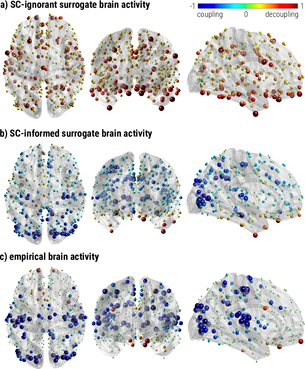
To assess the significance of this alignment, the authors generate two families of surrogate functional datasets. The “SC‑ignorant” surrogates preserve only the degree distribution of the structural graph, discarding any true anatomical information, while the “SC‑informed” surrogates retain the actual structural harmonics but randomize the sign of their coefficients at each time point. Comparing functional connectivity derived from these surrogates with empirical functional connectivity shows that SC‑informed surrogates reproduce many structural patterns but lack the contrast seen in real data, confirming that empirical FC reflects a non‑random combination of structural components.
The SDI is then defined per brain region as the log‑ratio of the energy contained in high‑pass (high‑frequency, structurally decoupled) versus low‑pass (low‑frequency, structurally coupled) filtered versions of the functional signal. Statistical significance is evaluated against the null distribution generated by the SC‑informed surrogates. Results reveal a clear gradient: primary sensory and motor cortices (visual, auditory, somatomotor) exhibit low SDI values, indicating strong structure‑function coupling, whereas association areas in the orbitofrontal, temporal, parietal, and prefrontal cortices display high SDI values, reflecting functional activity that is relatively independent of the structural scaffold. This gradient is highly reproducible across the two resting‑state sessions (spatial correlation r = 0.90 for empirical data, r = 0.99 for surrogates).
To link this neurobiological gradient to behavior, the authors perform a NeuroSynth meta‑analysis using 24 topic terms. The analysis shows that regions with low SDI are associated with low‑level processes such as multisensory perception, motor execution, and basic auditory/visual processing, whereas high‑SDI regions are linked to higher‑order functions including reward, emotion, social cognition, language, memory, and cognitive control. This functional hierarchy mirrors previously reported gradients based solely on functional connectivity, as well as gradients derived from cortical microstructure, gene‑expression profiles, and intrinsic timescale hierarchies.
In discussion, the authors argue that strong coupling in sensory‑motor areas likely reflects the need for rapid, reliable responses to external stimuli, whereas higher‑order cognitive processes benefit from a more flexible, less constrained functional repertoire. They suggest that the SDI provides a powerful framework for probing structure‑function relationships in health and disease, potentially revealing region‑specific decoupling patterns in neuropsychiatric disorders. Overall, the study demonstrates that the degree of structural‑functional coupling varies systematically across the cortex and aligns with a well‑established behavioral hierarchy, offering new quantitative tools for systems neuroscience.
Comments & Academic Discussion
Loading comments...
Leave a Comment